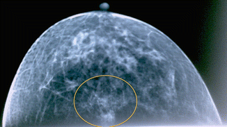
Fig. 20.1
Palpable mass in the left submammary fold, ultrasound, and mammography

Fig. 20.2
Palpable mass in the left submammary fold, ultrasound, and mammography
20.2 Surgery
Figure 20.3 shows preoperative drawings around the nipple for a 4 cm neo-areola. After sentinel biopsy revealed no cancer cells within the sentinel lymph node, the skin around the nipple-areola complex was deepithelialized (Fig. 20.4). The lower central segment was resected with the pectoralis fascia en bloc (Fig. 20.5a


Stay updated, free articles. Join our Telegram channel

Full access? Get Clinical Tree


