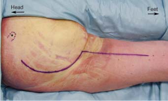
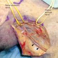
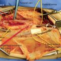
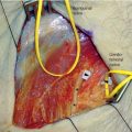
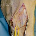
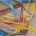
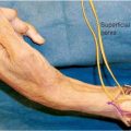
11 The ventral rami of the fourth lumbar to third sacral nerve roots unite to form the sciatic nerve. In the lesser pelvis the nerve lies anterior to the piriformis muscle. Just caudad to this muscle the nerve enters the buttock through the sciatic foramen. The nerve then moves laterally in an oblique direction beneath the gluteus maximus muscle toward the midline of the posterior aspect of the leg. On its medial side it is accompanied by the posterior cutaneous nerve of the thigh and the inferior gluteal artery. At a point midway between the ischial tuberosity and the greater trochanter the nerve turns downward over the gemelli muscles, the obturator internus tendon, and the quadratus femoris, and leaves the buttock to enter the thigh beneath the lower border of the gluteus maximus. The nerve descends in the thigh close to the midline, lying on the adductor magnus and being crossed obliquely by the long head of the biceps femoris muscle. In the majority of people the nerve divides into tibial and common peroneal branches at the middle to distal third of the thigh. In a small number of individuals the nerve may divide into its tibial and common peroneal branches as high up as the sciatic notch. In the buttock the nerve supplies an articular branch to the hip as well as vascular filaments to the inferior gluteal artery. In the thigh it supplies muscular branches to both heads of the biceps femoris muscle, semimembranosus, semitendinosus, and ischial head of the adductor magnus muscle. After intubation and induction of general endotracheal anesthesia on a stretcher, the patient is placed on the operating table in the prone position on abdominal bolsters. The arms are brought forward on arm boards. The entire ipsilateral buttock and leg down to the popliteal fossa are draped for surgery. If sural nerve grafting is anticipated, then the lower part of the extremities are also draped out. A curvilinear incision in a reverse question mark shape is fashioned.1 The stem of the question mark follows the midline of the posterior aspect of the thigh, whereas the curve follows the inferior margin of the gluteus muscle around the contour of the buttock and up onto the lateral aspect of the buttock (Fig. 11-1). The skin and subcutaneous tissue are divided after being infiltrated with lidocaine 1% with epinephrine in a 1:100,000 solution. The inferior margin of the gluteus maximus muscle is apparent from the fibers that travel in a medial to lateral direction (Fig. 11-2). At the midline of the leg, just inferior to the edge of the gluteal muscle, is the long head of the biceps femoris muscle (Fig. 11-3). Just lateral and deep to the long head a fat pad is found. The sciatic nerve is located within this fat (Figs. 11-4 and 11-5). The long head of the biceps femoris muscle crosses the path of the sciatic nerve from a medial to lateral direction. If a long exposure of the nerve in the thigh is required, the muscle must be mobilized so that it may be retracted as needed. The plane between the biceps femoris and the semitendinosus muscle must be developed (Fig. 11-6). When the plane is opened fully the nerve becomes visible with the biceps femoris lying laterally and the semitendinosus muscle medially (Fig. 11-7). Muscular branches to the biceps, semitendinosus, and semimembranosus from the sciatic should be preserved (Fig. 11-8). For even more distal exposure of the sciatic nerve the incision may be extended distally along the posterior midline of the thigh, tracing the nerve beneath the long head of the biceps femoris muscle and into its division into the common peroneal and tibial nerves (Fig. 11-9). For a more proximal exposure of the sciatic nerve the gluteus muscle must be divided. A cuff of muscle both medially and laterally must be left for reattachment at the completion of the procedure (Fig. 11-10). Once the muscle is divided, it is reflected in a unit medially for exposure of the sciatic nerve in the buttock (Fig. 11-11). For exposure of the nerve even more proximally the gluteus is divided higher up onto the buttock and the muscle retracted further medially (Fig. 11-12). The sciatic nerve may be traced up to the sciatic notch as it emerges from the pelvis. The most proximal portion is covered with the piriformis muscle, whereas the obturator internus and gemelli muscle lie beneath it (Fig. 11-13). Care must be taken close to the sciatic notch to avoid injury to the inferior gluteal artery. The transected artery may retract intrapelvically and require an intraabdominal approach to control resultant bleeding. Postoperative ischemic complications may also occur secondary to the arterial transection. Exposure of the sciatic nerve proximal to the sciatic notch requires an abdominal approach.
SCIATIC NERVE
ANATOMY
POSITIONING AND SURGICAL EXPOSURE
In Leg
In Buttock
Sciatic Nerve
Only gold members can continue reading. Log In or Register to continue

Full access? Get Clinical Tree


