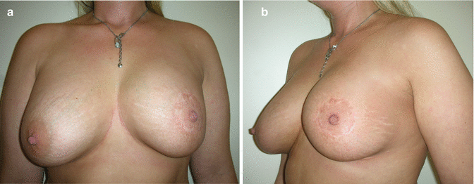
Fig. 55.1
(a, b) Preoperative view. The cancer is located periareolarly in the 12 o’clock position
The patient had a medium-sized breast with some ptosis, which was a little bit more pronounced on the left breast, and two scars in the inframammary fold (from the prior augmentation) (Fig. 55.1a, b).
55.2 Surgery
In order to correct the ptosis, quadrantectomy was done as a round block mammoplasty with the nipple elevated 2 cm on the breast median. Sentinel lymph node biopsy found two negative sentinel nodes.
55.3 Clinical and Cosmetic Outcome
The postoperative course was uneventful. Final histology found a 15 mm invasive cancer with the closest margin being 10 mm towards the major pectoralis muscle.
The patient underwent radiation treatment with a boost to the tumor bed and endocrine therapy was suggested for 5 years. The cosmetic result 1 year after surgery was rated as good by the surgeon and also the patient was very satisfied with the cosmetic outcome (Fig. 55.2a, b).
Get Clinical Tree app for offline access


