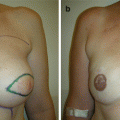
Fig. 13.1
Preoperative front and right oblique views with healing partial peri-areolar incision from previous excision
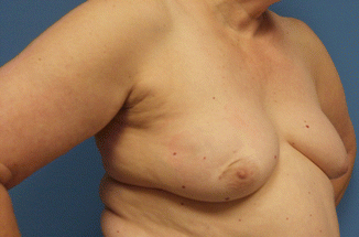
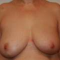
Fig. 13.2
Preoperative front and right oblique views with healing partial peri-areolar incision from previous excision
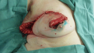
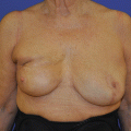
Fig. 13.3
Large inferio-medial rotational advancement flap
Related posts:
 Doughnut Lumpectomy: Caveat II
 Doughnut Lumpectomy: Caveat I
 Superior-Based Pedicle Quadrantectomy and Defect Reconstruction with Inferior-Based Pedicle: Secondary Prophylactic Mastectomy and Implant Reconstruction
 Segment Resection of a Breast Cancer in the Submammary Fold Using a Vertical Reduction Technique
 Correction of a Postlumpectomy Deformity Scar by Lipofilling
 Nipple-Areolar Complex (NAC) Reconstruction: Good Case
Stay updated, free articles. Join our Telegram channel

Full access? Get Clinical Tree


