Chapter 26. Rib Grafting
C. Spencer Cochran, MD; Jack P. Gunter, MD
INDICATIONS
Although septal cartilage is generally considered the preferred grafting material in rhinoplasty, severe deformities or a paucity of available septal cartilage requires alternative sources of grafting material. This is particularly true in patients who require dorsal augmentation and in secondary rhinoplasty patients with structural deformities resulting from previous procedures.
PREOPERATIVE PREPARATION
The choice of which rib to harvest depends on the planned use and grafting requirements, because the amount of cartilage required dictates whether the cartilaginous segment from one rib, rib and a portion of another, or the entire cartilage segments of 2 ribs need to be harvested. The surgeon should choose the cartilaginous portion of the fifth, sixth, or seventh rib that provides a straight segment long enough to construct all required grafts. If additional grafts are needed, a part or the entire cartilaginous portion of an adjacent rib may be harvested.
In older patients, ossification of the cartilaginous ribs can make graft preparation difficult and preclude its use. A preoperative CT scan of the ribs and sternum can help delineate the extent of calcification in patients in whom there is a high index of suspicion. In this case, some surgeons recommend diced cartilage within a temporalis fascia wrap.
ANESTHESIA
General anesthesia with endotracheal intubation is preferred for this procedure. This allows the patient to be completely still for the duration of the procedure and allows for a protected airway.
POSITION AND MARKINGS
The patient is placed in the supine position. After adequate anesthesia is administered, the patient is prepped and draped in a sterile manner. In female patients, the incision is marked slightly above the inframammary fold and measures 5 cm in length (Fig. 26-1). In males, placement of the incision is not as important unless there is a hair-bearing area in which the incision can be camouflaged. If not, the incision is usually placed directly over the chosen rib to facilitate the dissection. Although rib cartilage can be harvested from either the patient’s right or left side, harvesting rib cartilage from the patient’s left side allows for a 2-team approach.
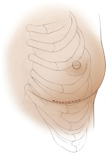
Figure 26-1 The skin incision (red line) for harvesting rib cartilage in female patients is placed parallel and slightly superior to the inframammary fold and measures approximately 5 cm.
DETAILS OF PROCEDURE
The skin is incised with a scalpel, and the subcutaneous tissue is divided with electrocautery. Once the muscle fascia has been reached, the surgeon palpates the underlying ribs and divides the muscle and fascia with electrocautery directly over the cartilaginous portion of the chosen rib (Fig. 26-2). The rib is exposed from its sternal attachment medially to the bony-cartilaginous junction laterally. Identification of this junction is facilitated by the subtle change in color at the interface: The cartilaginous portion is generally off-white, whereas the bony rib demonstrates a distinct reddish-gray hue.
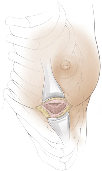
Figure 26-2 After incising the skin, dissection is carried out down through the subcutaneous tissue, and the surgeon divides the muscle and fascia with electrocautery directly over the cartilaginous portion of the chosen rib.
After exposing the selected rib, an incision is made through the perichondrium along the long axis of the rib (Fig. 26-3
Related posts:


Stay updated, free articles. Join our Telegram channel

Full access? Get Clinical Tree


