Fig. 17.1
Deep ulcer with necrotic eschar was found on the medial malleolus in patient with RA
Fig. 17.2
(a) Right finger necrosis due to ischemia was found in patient with RA. (b) Angiography showed the obstruction of digital artery due to vasculitis of medium-peripheral vessel
Pathologic features of rheumatoid vasculitis include mononuclear cells or neutrophilic infiltration of the vessel wall of small and medium vessels.
17.2.2 Neutrophilic Dermatoses
Neutrophilic dermatoses are the conditions that have an inflammatory infiltrate consisting of mature polymorphonuclear leukocytes with no evidence of infection; these include Sweet’s syndrome and pyoderma gangrenosum. The pathogenesis of neutrophilic dermatoses is believed that these disorders represent a state of altered immunologic reactivity because they generally respond to systemic glucocorticoids and other immunomodulatory therapies [1].
17.2.3 Venous Stasis
Ulcerative lesions may also result from venous stasis. Ankle joint dysfunction caused by RA reduces ankle movement, which is responsible for impairment of the normal venous pump function and leads to venous hypertension [3]. This unfavorable state leads to increased tissue fibrosis and decreases the diffusion of oxygen to the skin, producing skin fragility and results in venous ulceration [1] (Fig. 17.3). Leg ulcers in RA are associated with venous disease in as many as 45 % [8].
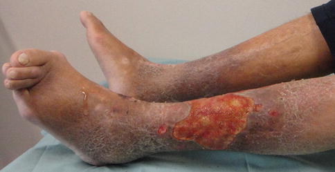
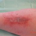
Fig. 17.3
Venous stasis ulcer located at the lower leg surrounded by varicosis. Toe and ankle deformities caused by RA were also found
17.2.4 Arterial Disease
Arterial insufficiency is one of the reasons to develop ulcer, especially, in the toes or feet of patients with RA and several connective tissue diseases, such as systemic sclerosis and scleroderma. Pun et al. found arterial insufficiency in 36 % of ulcerated legs in 26 patients with RA, and Baker et al. did found ischemia in 41 % of those in 27 patients with RA [8].
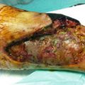
17.2.5 Corticosteroid Therapy
Medications used to treat RA can cause skin changes [4]. It is a common clinical practice to use systemic glucocorticoids to suppress RV and other connective tissue diseases. Glucocorticoids are administrated to about half of patients with RA leg ulcers. Effects of glucocorticoids inhibiting wound healing include: stabilization of lysosomal membranes which inhibits the release of chemical mediators, suppression of fibroblasts and immunity, and inhibition of collagen fiber synthesis causing skin atrophy [5]. Atrophic skin of RA patients treated with continuous glucocorticoids can be easily torn and develop lacerations, which are likely to be more severe because infections are common (Fig. 17.4).
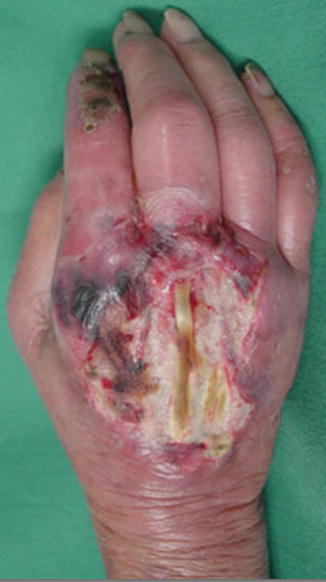
Fig. 17.4
Small wound of the hand of SLE patients has become more severe because infections occurred
17.3 Ulcers in Other Connective Tissue Diseases
17.3.1 Systemic Lupus Erythematosus (SLE)
Ten to twenty percent of patients with SLE develop cutaneous vasculitis and show purpuric papules, which sometimes cause ulceration [8]. The lower extremity is a common site and leg ulcer caused by leukocytoclastic vasculitis or necrotizing arteritis was found in 5–6 % of patients with SLE (Fig. 17.5).
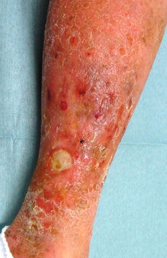
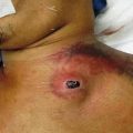
Fig. 17.5
A patient with SLE had a leg ulcer for 2 years
17.3.2 Systemic Sclerosis
Sclerodermatous change develops usually in the feet and legs, which sometimes results in ischemic necrosis and ulceration of the toes. Early skin changes in systemic forms may include edematous change, which lasts or is replaced by thickening and tightening of the skin. Painful ulcerations appear, especially on the area of the skin overlying bony prominence. These lesions are hard to heal [4].
17.3.3 Dermatomyositis
A small-vessel vasculitis and calcinosis in the subcutaneous tissue cause ulceration usually on the feet. Healing is slow and ulcer often requires debridement of calcinotic material surgically [4].
17.3.4 Sjögren’s Syndrome
Sjögren’s syndrome is associated with RA, SLE, and some inflammatory connective tissue disease. Cutaneous magnifications include Raynaud’s phenomenon (in 33 % of patients), dryness of skin, purpura, and vasculitic ulcers of the legs [4].
17.3.5 Scleroderma
Although scleroderma (SSc) is a clinically heterogeneous disorder, the loss of cutaneous elasticity and accompanying tightness followed by thickening and hardening of the skin is an almost universal manifestation. Painful digital ulcers that occur on the fingertips as a result of local ischaemia and vascular insufficiency are a frequent complication. While some SSc patients have skin lesions that remain largely confined to the extremities, others exhibit skin thickening that extends progressively from the extremities to the trunk [4].
Related posts:

Stay updated, free articles. Join our Telegram channel

Full access? Get Clinical Tree


