Revision Total Hip Replacement

CHAPTER 34 Revision Total Hip Replacement


Posterior Approach


The posterior approach to the hip is a versatile and extensile approach that can be used effectively for the majority of hip revision procedures. The incision is typically curvilinear, extending from the posterior superior iliac spine (PSIS) toward the tip of the greater trochanter, and then extending distally along the shaft of the femur as far as required to meet the goals of the operation. The distal extension is determined by factors such as the need to remove an existing stem, locate a cortical defect or fracture, perform an osteotomy, or apply a bone graft.


INDICATIONS AND CONTRAINDICATIONS


The posterior approach is indicated when access to the femur is a primary requirement for reconstruction. The posterior approach can provide visualization of the entire lateral and anterior femoral cortex distally, as well as the proximal femoral cortex and femoral canal. Likewise, the posterior approach is appropriate for wide acetabular exposure that can also expose the posterior column and superior rim defects that do not extend far into the quadrilateral surface of the pelvis or into the anterior column. The posterior column is accessible for most indications, including plating, hardware removal, sciatic nerve visualization, placement of a jumbo spherical cup or acetabular cage, and bone grafting.


The limitations of and contraindications to the posterior approach are primarily based on the lack of access to the anterior column and the quadrilateral surface of the pelvis or on the need to avoid operating through the posterior capsule of the hip joint. The proximal extension of the posterior approach is limited by the lack of excursion of the superior gluteal neurovascular pedicle emerging from the sciatic notch and supplying the gluteus medius muscle. This pedicle cannot be stretched sufficiently to allow a wide exposure of the lateral wall of the pelvis without risking denervation of the gluteus medius and tensor fascia lata muscles. When the integrity of the posterior hip capsule is of primary importance because of considerations of joint stability, the posterior approach is not the approach of choice owing to the risk of posterior hip dislocation.


PREOPERATIVE PLANNING


Preoperative planning starts with determining whether the posterior approach will afford access to that part of the pelvis and femur critical to meet the reconstructive goals. The posterior approach will allow access to the superior rim of the acetabulum, the acetabulum itself, contained anterior column defects, any type of posterior column defect, and virtually the entire femur. The posterior approach is not ideal for large superior or anterior pelvic or anterior column defects. With that anatomic footprint in mind, careful imaging will allow a determination of whether the posterior approach is the right choice in a given situation.


Other preoperative considerations include prior incisions and procedures, a history of posterior instability, or retained hardware. Although the posterior approach can be used after any prior approach, it is advisable to use prior incisions or approaches when there is no advantage to using the posterior approach. Examples might include the prior use of an anterior approach or an anterolateral approach for liner exchange, débridement, or isolated acetabular revision (particularly with a history of posterior instability).


Once it has been determined that the posterior approach is appropriate with regard to skeletal access, further consideration should be given to the length of the incision required and the exact placement of the incision. Procedures that require a more extensive exposure of the posterior column can be facilitated by placing the incision more posteriorly. Situations in which a trochanteric osteotomy is contemplated might be better served by a straighter, more trochanteric-based incision. A more distal extension of the approach provides access to the femoral shaft when required.


TECHNIQUE


After the surgical site is marked, the patient is positioned in the lateral decubitus position. All downside pressure points should be padded, with particular attention paid to the peroneal nerve just below the fibular head on the downside leg. An axillary roll under the thorax provides protection to the downside shoulder and the brachial plexus. For procedures that are anticipated to take longer or that may be associated with larger volumes of blood loss because of an extended exposure, appropriate monitoring of fluid status with central pressure, a urinary catheter, and use of an intraoperative blood salvage device should be considered. The leg is draped free with the foot isolated by an impervious drape so that the leg can be manipulated during surgery. Adhesive drapes obviate the need for wound towels and can isolate any exposed skin surface, which is a potential source of contamination if exposed during the procedure.


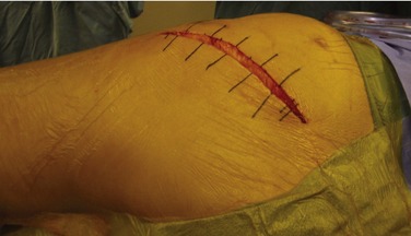
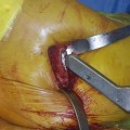
Planning the skin incision entails the use of surface landmarks such as the PSIS, the sciatic notch, the tip of the greater trochanter, and the femoral shaft. The incision will be centered over the greater trochanter, with the proximal limb extending toward the PSIS, and the distal limb paralleling the femoral shaft (Fig. 34-1). The skin is incised with a knife, and the initial dissection is carried through the subcutaneous fat down to the tensor fascia. The fascia may be exposed by carefully elevating the fat just enough to expose the edges of the fascia for later closure. The fat and skin should be handled with care throughout the surgery to prevent devascularization or injury that could predispose to wound dehiscence or breakdown of the skin closure.


The deep dissection passes through the gluteus maximus (innervation—inferior gluteal nerve), posterior to the gluteus medius muscle (innervation—superior gluteal nerve) and gluteus minimus muscle (innervation—superior gluteal nerve), and through the insertion of the piriformis muscle, the superior gemellus, the obturator internus, the inferior gemellus, the obturator externus, and the quadratus femoris muscle in that order from proximal to distal (innervation—sacral plexus, L5, S1, S2).

Stay updated, free articles. Join our Telegram channel

Mar 10, 2016 | Posted by in Reconstructive surgery | Comments Off on Revision Total Hip Replacement

Full access? Get Clinical Tree

Get Clinical Tree app for offline access