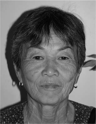
5 Differences between the Asian and Caucasian face and neck are manifested in skin thickness and texture, patterns of fat accumulation, and, perhaps most importantly, skeletal structure. When analyzing these anatomic differences, it is important to note that just as in the Caucasian population, considerable individual variations in anatomy exist among Asians, perhaps the most important variable being related to the geographic latitude of origin. In general, Asian skin tends to be thicker, and the face and neck tend to accumulate more fat during the aging process than the Caucasian. Prominent malar eminences accompanied by premaxillary retrusion and a relatively wide mandible combine to generate a more square, less angular configuration. A combination of heavier soft tissue and a flat facial skeleton translates into less resistance to gravitational forces with aging. Although the classical Western description of Asian skin having a yellowish coloration is not without some biological justification, there is actually a wide variation in the color and texture of the skin. Generally, skin pigmentation is darker in Asians originating from southern latitudes, whereas individuals from northern areas tend to display light-colored, somewhat milky skin pigmentation. The yellowish tint of Asian skin is largely a consequence of the number and distribution of melanocytes rather than of variations in the skin lipoproteins. As is the case with skin color, a wide variation in texture and thickness is apparent among Asians, the dermis tending to be more fibrous (or dense) in individuals with darker pigmentation. The skin of lightly pigmented Asians, however, is generally thicker and denser than of equivalently pigmented Caucasians. Greater collagen density is manifested in a tendency toward a more vigorous fibroplastic response toward wound healing, which may produce hypertrophic scarring and prolonged hyperemia during scar maturation, an incidence worthy of note even in lightly pigmented Asians (especially with regard to prolonged incisional hyperemia). Although I cannot recall ever having observed a true keloid in an Asian patient following cosmetic surgery, there is definitely an increased incidence of hypertrophic scar formation in the postauricular region, at the junction of the superior helical and temporal portions of rhytidectomy incisions, and in the medial canthal area. Curiously, hypertrophic scars are extremely rare in the alar crease. Because of differences in dermal thickness, the Asian face undergoes photoaging differently than the Caucasian face, the Asian face tending to exhibit fewer fine wrinkles than a Caucasian of comparable age. The Asian face, however, shows a higher incidence of pigmented dermatoses (e.g., seborrheic keratoses, lentigines, and other melanocytic lesions) (Fig. 5-1) with aging, and fair-complexioned Asians often exhibit rhytidosis comparable to Caucasians of a similar age (Fig. 5-2). Figure 5-1 Lentigos in a 35-year-old Asian woman. Skin flaps developed during cervicofacial rhytidec-tomy are more tolerant of tension; however, this is counterbalanced by an increased incidence of hypertrophic scar formation, as noted above. Some clinicians have described a more vertical orientation of subdermal vascularity in the Asian face and suggest that this anatomic pattern might reduce collateral circulation in skin flaps. I have noted no such differences in vascularity and conclude that such suggestions are of no clinical significance with regard to the tolerable extent of undermining. During the aging process, the Asian face typically accumulates greater volumes of fat than its Caucasian counterpart. This fat accumulation tends to concentrate in the jowl, nasolabial mound, and buccal regions. The clinical significance of fat accumulated in these areas as accentuated by the skeletal structure of the Asian face has important implications for successful surgical management of facial aging. Figure 5-2 Extensive rhytidosis in 59-year-old Asian woman. Fat accumulation in the neck that contributes to the so-called double chin is less common in Asians younger than 40 years old than in comparably aged Caucasians; submental fat accumulation, however, occurs more frequently in the Asian than in the Caucasian neck after age 40. Although the platysma is generally thicker in the Asian neck, the incidence of diastasis is considerably less, occurring in ~30% of patients who undergo cervicofacial rhytidectomy (Fig. 5-3), as compared with 60% of Caucasians undergoing this procedure. Perhaps the most significant anatomic differences between the Caucasian and Asian face are related to skeletal structure. As noted above, the Asian face is characterized by prominent malar eminences associated with relative deficiency of the premaxillary region that results in shallowness of the midfacial region. Wide, prominent mandibular angles are often present, contributing to a square, flattened face. Microgenia may be somewhat more common than in the Caucasian, but the cervical deformity attributed to a “low hyoid” bone is distinctly less common in the Asian neck. Figure 5-3 Diastasis of the anterior platysma. The basic subcutaneous face lift operation was described by Lexer in 1916, and, perhaps surprisingly, except for the extent of undermining, no substantial improvements or modifications in the basic procedure were introduced until the 1970s. Since that time there has been a vast proliferation of variations and modifications of the face lift operation; thus, not surprisingly, considerable controversy exists regarding the optimal surgical approach for facial rejuvenation. A brief historical survey of the evolution of face lift techniques is instructive and valuable in assisting the surgeon in formulating an approach to the aging face and neck. The first such innovation was the Skoog technique introduced in 1973.1 This procedure consisted of elevation of a platysmal flap in the neck and lower face without dissection of the overlying skin. In 1976, Mitz and Peyronie2 described the muscle and fascial complex of the lower face, which they termed the superficial musculoaponeurotic system (SMAS). Surgeons quickly became enamored with SMAS techniques, and numerous variations have been described (limited SMAS, lateral SMAS, conventional SMAS, extended SMAS, and anterior SMAS procedures); however, for the most part, the only actual modifications relate to the extent of flap elevation and/or the direction of flap suspension. Experience has suggested that following any SMAS dissection, improvement is generally limited to the lower face (i.e., the jowl and mandibular regions). Incorporation of platysmal elevation and repositioning in conjunction with SMAS dissection extends improvement into the cervical region but does not improve the nasolabial folds, resulting in what some surgeons term “facial disharmony.” Of significance in assessing the role of SMAS dissection in cervicofacial rhytidectomy is the fact that the precise anatomy and clinical importance of this structure are currently matters of controversy, a situation that I term “SMAS confusion.” Jost and Levet3 claim that the anatomic interpretations of Mitz and Peyronie are erroneous, the SMAS layer that they described being merely a superficial fascial layer that lacks sufficient tensile strength to hold plication sutures. According to their anatomic studies, Jost and Levet state that the true SMAS is actually a deeper structure contiguous with the parotid fascia that necessitates actual exposure of the lobular structures of this gland for proper dissection. Embryologic considerations, as well as histologic findings of muscle fibers in this tissue, support labeling of the true SMAS as a vestige of the platysma aponeurosis. Regardless of its precise anatomy, it is important to consider that the SMAS itself does not participate in facial ptosis, and thus any role that it may play in facial rejuvenation is limited to service as a vehicle to assist in repositioning of more anterolateral (or superficial) ptotic tissues, being of value by diverting tension from the skin flap. Efforts to improve the nasolabial fold were largely unsuccessful until 1988, when Hamra4 described the deep plane rhytidectomy. This procedure involved elevation of the malar fat pad following its dissection from the superficial surface of the zygomatic musculature. This dissection was accompanied by elevation of a SMAS flap in the lower face, and was the first technique that allowed true anatomic repositioning of cheek fat in an effort to efface the nasolabial folds. Not completely satisfied with results of this procedure because, although the nasolabial fold did demonstrate improvement, stigmata of aging in the superior aspect of the malar region and the lower portion of the orbicularis muscle persisted, Hamra5 enhanced the deep plane rhytidectomy by incorporating dissection of orbicularis oculi into the flap, producing a bipedicled musculocutaneous flap based on the platysma and orbicularis blood supply. This operation was termed composite rhytidectomy, and a key concept of this procedure involved the ability to suspend the flap under “extraordinary tension” to efface the nasolabial fold. Hamra maintained that the composite rhytidectomy was the only anatomically correct method for deep plane manipulation, and that, in contrast, SMAS dissection disrupted the normal relationships among the orbicularis, cheek fat, platysma, and skin. Of major interest in this regard was a study published in 1996. Ivy, Lorenc, and Aston6 compared various SMAS procedures and composite rhytidectomy, different procedures being performed on contralateral sides of the face in 21 patients. The surgeons reported that on the operating table, composite rhytidectomy produced more dramatic effacement of the nasolabial folds as compared with limited SMAS dissection, but that this difference disappeared within 24 hours postoperatively, and no difference was observed by a panel of three surgeons or the patients themselves on evaluation 6 and 12 months postoperatively. Following long-term examination of his results, Hamra7 stated that neither the deep plane nor early versions of the composite rhytidectomy resulted in substantial lasting effacement of the nasolabial fold (in contrast to the effect of SMAS dissection that yielded long-term improvement in the lower face), and that the only method of producing long-term improvement was direct excision (nasolabioplasty). He further suggested that an improved composite rhytidectomy incorporating superomedial repositioning of the orbicularis-zygomatic muscle reduced the incidence of postrhytidectomy facial disharmony that results from disparity between the longevity of SMAS dissection (lower face) and midfacial rejuvenation. To summarize, SMAS dissection tends to improve results in the jowl and mandibular regions, but it is associated with relatively poor nasolabial effacement, a disparity that may produce “facial disharmony.” One possible reason for this effect is the fact that SMAS may actually result in tethering of the zygomatic muscles, dissipating “lifting” forces applied intraoperatively by plication or suspension. Of importance is the fact that many surgeons feel that, in spite of the initial optimism surrounding the deep plane and composite face lifting techniques, there is little evidence of lasting benefit as compared with extended subcutaneous lifting. Other surgeons approached the problem of midfacial ptosis utilizing subperiosteal dissection inspired by the techniques of Tessier, which were developed in his epic contributions to reconstruction of craniofacial deformities. In 1988, Psillakis and his associates8 described a subperiosteal approach for which they claimed enhanced results but which was associated with a high incidence of facial nerve injury. Ramirez et al have been the most persistent disciples of the subperiosteal face lift, first describing this technique in 19919 and stating that their modifications reduce the incidence of facial nerve complications from 20 to ~2%. The subperiosteal approach described by Ramirez et al begins with a coronal incision, thus allowing subperiosteal dissection to the level of the superior and lateral orbital rim. Laterally, the dissection continues deep to the temporoparietal fascia; at a point ~3 cm above the zygomatic arch, dissection is deepened through both layers of the temporalis fascia, continuing to the superior aspect of the zygomatic arch, at which point the deep temporal fascia and periosteum of the entire zygomatic arch are incised on its posterior surface with a scalpel. The subperiosteal dissection is connected with the superior and lateral orbital rim dissection plane, and dissection over the anterior surface of the zygomatic arch and body continues over the maxilla inferiorly to the pyriform aperture. Dissection is continued inferiorly over the masseter muscle, this freeing of the cheek tissues from the masseter being the key factor in allowing midfacial elevation. Currently, there is much enthusiasm (although I sense that it is waning) for endoscopic,10–12 transble-pharoplasty, 13,14 and transbrow15 approaches to the midface. (Many such techniques depend on suture or “sling”(utilizing alloplastic or autogenous material) suspension of musculofascial tissues as a vehicle for skin-fat elevation.16–18 Although I certainly applaud all attempts at innovation and technical improvement, I maintain a cautious attitude, given the surgical community’s experience with enthusiastic embracement of previous techniques that all too often failed to yield the long-term benefits suggested by early results. At any rate, even the enthusiasts for such procedures suggest that they are most effective for younger patients who exhibit minimal redundant tissue. Given the tendency for the aging Asian face to accumulate large volumes of fat in the central cheek region, I feel that particular caution is warranted in application of such techniques to this population. As might be expected, advocates of deep approaches to the midfacial tissues have been vigorously opposed by other surgeons concerned by the high incidence of prolonged edema, facial nerve injury, and associated complications. Several experienced surgeons have suggested that the pendulum has swung too far, recommending, in the interest of patient safety, as well as the lack of definitive evidence proving long-term efficacy, a return to more traditional techniques of facial rejuvenation. In spite of this tangled web of controversy regarding the optimal surgical approach (that remains uncertain and in a state of continuing evolution and discovery), most surgeons do agree that regardless of the techniques employed, the goals of rejuvenation should be the same: a natural, long-lasting appearance associated with a relatively rapid healing period and a low incidence of complications. Each individual surgeon has strong opinions regarding the relative importance of various facial components and the most efficacious method of achieving optimal results. Most, however, agree that achieving these goals requires repositioning of ptotic skin as well as some sort of manipulation (not necessarily dissection and/or elevation) of deeper structures. A major problem in face lift surgery relates to the fact that cosmetic surgery is not an exact science. Results are difficult to quantitate with scientific precision. Double-blind studies are impractical because of both ethical and logistical considerations, and reliable animal models are not available. A major reason for absence of proof of long-term efficacy is the fact that most satisfied patients do not return for extended follow-ups. Most individual surgeons are convinced, largely for anecdotal reasons, that their techniques produce the best results. Finally, the “herd instinct” seems to be particularly powerful in cosmetic surgery. As a most interested observer and a participant in this continuing 30-year drama, my preferences have undergone significant metamorphoses as a function of time. I have chosen my current rhytidectomy techniques (which, of course, are subject to change as clinical evidence evolves) based on the following observations and convictions: Surgeons are understandably frustrated by the current state of affairs in cervicofacial rhytidectomy. To some extent, public relations agents have replaced peer review, “innovations” are hyped for their marketing value, and as a result many patients demand the latest techniques that lack proof of efficacy. Responsible surgeons recognize that rapid healing and restoration of social and occupational functioning as well as safety are important factors in patient satisfaction. In many respects, these factors exceed the importance of total eradication of all vestiges of facial aging, especially when more complex techniques involve prolonged healing and an increased incidence of complications. The primary techniques that I employ for cervical facial rhytidectomy are described in the following sections. As noted above, since the description of SMAS in 1976, surgeons have attempted to devise an enhanced, longer lasting face lift based on dissection of the deep facial and cervical tissues. Although mobilization and modification of deep cervical structures (i.e., platysma muscle) have become accepted practice for most surgeons, many have been reluctant to incorporate the aggressive dissection of deeper facial structures into their surgical routines because of the seeming complexity of these procedures coupled with the increased risk of facial nerve injury (as well as prolonged facial edema). In addition, the initial enthusiasm for SMAS mobilization has waned somewhat, as it has become apparent that, although such techniques do enhance effacement of the jowl, little or no long-term improvement is noted in the midfacial (i.e., nasolabial mound and fold) regions following even the most extensive SMAS dissection. New insights into the pathophysiology of midfacial ptosis have clarified understanding of the failure of SMAS dissection to rejuvenate this region. Because midfacial aging is largely a consequence of ptosis of the malar fat pad (not accompanied by ptosis or diminished tone of the mimetic musculature), in some cases associated with localized hypertrophy of cheek fat, rejuvenation of this area requires direct elevation (or other modification) of the ptotic malar fat pad and overlying skin.19 Although this goal can be achieved via supra- or subperiosteal approaches, I initially felt that the subperiosteal approach provided a direct route associated with a lower incidence of complications than supraperiosteal dissection.
Rejuvenation of the Aging Asian Face and Neck
Although the goals of rejuvenating the aging face and neck are the same in the Asian as in the Caucasian patient, certain anatomic differences, as well as biological manifestations of the aging process, are noteworthy, as they translate into variation in surgical approach to cervicofacial rhytidectomy.
♦ General and Anatomic Considerations
Skin

Fat

Platysma Muscle
Skeletal Structure

♦ Formulating an Approach to Rejuvenation of the Aging Face and Neck
♦ Rhytidectomy (Full Face Lift)
Stay updated, free articles. Join our Telegram channel

Full access? Get Clinical Tree


