Fig. 27.1
A right leg ulcer in a 20-year-old male that originated from initial abrasion. He has no systemic disease
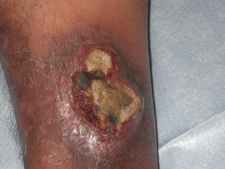
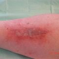
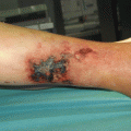
Fig. 27.2
The ulcer expanded with a purulent and necrotic base even though ointment was used
Pathergy is a specific but not sensitive finding of PG. The lesion sites expand radically, especially if the borders of the lesion site are traumatized by debridement or by other mechanical trauma [3]. No laboratory finding is diagnostic of PG. PG has no definite diagnostic criteria and is a diagnosis of exclusion. The diagnosis of PG is very difficult and based primarily on the clinical history, clinical manifestation, and biopsy result while being careful about misdiagnosis (Table 27.1). [1, 2, 6]
Table 27.1
Causes of ulcers mimicking PG
Infection |
Fungal |
Mycobacterial |
Necrotizing fasciitis |
Vascular occlusive disease |
Antiphospholipid-antibody syndrome |
Venous stasis ulceration |
Vasculitis |
Wegener’s granulomatosis |
Polyarteritis nodosa |
Neoplasms |
Lymphoma |
Leukemia cutis |
Drug reactions |
Hydroxyurea induced ulcer |
27.4 Treatments
If the patients have a systemic disease, the systemic disease should be preferentially treated. It is difficult to get cured completely by local wound management alone. Topical treatments are chosen depending on the purpose such as the prevention of secondary bacterial infection or the promotion of reepithelialization. Some topical agents such as tacrolimus, strong corticosteroids, and cyclosporine have reported efficacy in small case series. PG is commonly treated with systemic corticosteroids and/or cyclosporine [7].
Related posts:


Stay updated, free articles. Join our Telegram channel

Full access? Get Clinical Tree


