Intense Pulsed Light (IPL) is a light-emitting system that is capable of emitting filtered polychromatic broad bandwidth wavelengths between 515 and 1,200 nm.
The emission of wavelengths is controlled by both an internal filter that blocks undesired wavelengths from being emitted and a “heat-sink” effect that allows the controlled transfer of thermal energy from an object at high temperature to an object at lower temperature.
Introduction
Intense pulsed light (IPL) is a non-coherent filtered flash lamp capable of emitting a polychromatic broad bandwidth of light from 515 to 1,200 nm (approximately). It was first introduced in clinical studies in 1994 and cleared by the US FDA in late 1995, mainly for the treatment of leg veins. The device that first received approval was PhotodermTM (ESC/Sharplan, Norwood, MA-now, Lumenis, Santa Clara, CA). This technology is now marketed as the gold standard for treatment of signs of photoaging and its other recognized indications are treatment of virtually all skin vascular conditions, scarring, pigmentation, Poikiloderma of Civatte and hair removal.
The IPL Device
The IPL device consists of a flashlamp housed in an optical treatment head with water cooled reflecting mirrors. An internal filter overlying the flashlamp prevents wavelengths less than 500 nm from being emitted. In certain devices, cut-off filters of various wavelengths (515, 550, 560,570, 590, 615, 645, 690, 755 nm) are available which are placed over the window of the optical treatment head to eliminate other wavelengths. Other devices required the changing of the IPL head to manipulate wavelengths. In some of the optical heads the water circulates around the flashlamp and helps reduce the recycling times. A water based gel is used for the purpose of allowing optimal light transmission to the skin by decreasing the index of refraction of light. Also, it creates a “heat-sink” effect. Heat sinks function by efficiently transferring thermal energy (“heat”) from an object at a relatively high temperature to a second object at a lower temperature with a much greater heat capacity. This rapid transfer of thermal energy quickly brings the first object into thermal equilibrium with the second, lowering the temperature of the first object, fulfilling the heat sink’s role as a cooling device.
Bio Physical Interactions
Intense Pulse Light (IPL) systems function on the principle of selective photothermolysis by targeting specific ranges of wavelength. Small spot sizes used during treatment with IPL are associated with more scattering, yet heat distribution is better assimilated. In contrast, larger spot sizes have less scattering and are better targeted, but require greater thermal energy. A water-based gel filters out unnecessary wavelengths, such as near-infrared wavelengths that may be more damaging to the tissue. Tissue temperature is controlled by allowing thermal relaxation time (TRT) by a factor of ε = 2.72 between treatment sessions to prevent the elevation of epidermal temperatures above 70°C. |
Wavelength
IPL, like lasers, works on the principle of selective photothermolysis, first described by Anderson and Parrish in 1983.9 The target structures may have different absorption maxima such as hemoglobin absorbs at a wavelength of 580 nm while melanin has a spectral range of 400–750 nm. Longer wavelengths are absorbed lesser by the melanin and penetrate deep, thus reaching deeper blood vessels and avoiding epidermal damage. The bandwidth of IPL is modified by application of filters which exclude the lower wavelengths. In that spectrum, during a 10 ms pulse, relatively high doses of yellow light at 600 nm are emitted and other wavelengths are emitted much less.4 This wavelength facilitates selective absorption by bright red superficial vessels.
Vascular lesion treatment filters require filters such as 515, 550, 560, 570 and 590 nm. Hair removal can be performed using longer filters such as 615, 645, 695 and 755 nm. These filters can also be used to cause fibroblast stimulation.
Spot Size
Spot size along with wavelength affects penetration depth. Smaller the spot size, more rapid the scatter and more rapid the decay of fluence by depth (Table 1). Thus larger spot size would result in greater amount of energy being delivered to the skin. The light emitting planar surface of the IPL device functions through a water-based interface between the crystal and the skin. This water based gel, as mentioned earlier has functions of enhancing optical coupling, minimizing reflections, acting as a “heat sink” and maintaining continuity of index of refraction of the skin/air interface. The gel also helps filter higher wavelengths, thus removing some of the ineffective or potentially tissue damaging near-infrared wavelengths before being absorbed by the target. In order to treat deeper or larger vessels, the fluence can be modified and increased and the gel can be chilled to protect the overlying epidermis. A 1–2 mm layer of chilling gel can be used between the crystal and the skin when working with a large footprint of IPL, as has been termed as “floating” the crystal. This is especially useful when an integrated chilled crystal is not being used.
Table 1
Spot size vs. wavelength
Spot size | 1 mm | 2 mm | 5 mm |
|---|---|---|---|
Light penetration depth (595 nm) | 0.8 mm | 1.1 mm | 1.25 mm |
Light penetration depth (800 nm) | 1.5 mm | 2.0 mm | 2.5 mm |
Pulse Duration
Between two pulses, some amount of time is allowed for the epidermis to cool down and is referred to as thermal relaxation time (TRT). More accurately defined TRT is the amount of time taken for the temperature of a tissue to decrease by a factor of ε = 2.72 as a result of heat conductivity. TRT helps to prevent the elevation of epidermal temperatures above 70°C and prevents epidermal damage.
Epidermal thickness of 100 μ has a TRT of about 1 ms, a typical vessel of 100 μ has a TRT of approx. 4 ms. This can be used to predict that vessels greater than 300 μ would have a TRT of more than 10 ms and thus these vessels will cool more slowly than the epidermis, with a single pulse. For larger vessels, multiple pulses prove more advantageous with 10 ms or more delay times in between the pulses for epidermal cooling. The treatment of darker skin individuals (Fitzpatrick IV–VI), and/or patients with hyper-reactive melanocytes should be performed very carefully, best by using a 755 nm filter with a pulse delay of 50–100 ms, to allow sufficient cooling time for the epidermis. When treating leg veins, better results have been obtained with longer pulse duration (up to 50 ms).10
A study conducted to understand the principles of “double or multiple pulsing” using a 585 nm yellow dye laser on larger vessels of Port wine stains, showed that these vessels absorbed greater energy fluences before reaching purpura after double pulses spaced 3–10 ms apart.11 After a pump pulse delivering 80% of the fluence necessary for causing purpura, the fluence of a second probe pulse necessary to cause purpura was determined and was found to increase the interval between the two pulses, in a manner consistent with the thermal diffusion theory. For a small vessel (0.3 mm), heat distribution was assumed to occur instantaneously. For a larger vessel, heat had to pass from the inside of the superficial vessel wall and through the vessel to the deeper wall. Larger vessels required longer cooling time periods for the accumulated heat inside the core. These principles are effective when using a double pulse 585 nm yellow dye laser in the treatment of larger vessels of PWS (larger than 0.1 mm). After double pulses spaced between 3 and 10 ms apart, larger vessels absorbed greater energy fluences upon reaching purpura.11
A study using pulsed laser irradiation at 585 nm with short pulse (0.45 ms) and long pulse (10 ms) durations showed that long-duration pulses caused coagulation of the larger diameter vessels. Small caliber vessels and capillaries demonstrated resistance to photothermolysis.12 The use of IPL with extended pulse durations from 12–50 ms has caused larger vessels (0.5 mm or greater) to undergo more clinical photothermal coagulation while sparing the epidermis.6
Indications
Leg vein treatment
Hair removal
Vascular skin conditions
Scarring
Pigmentation
Poikiloderma of Civatte
Photorejuvenation
A two peak approach of dual short and long wavelengths is an effective method of treating the variably colored, multiple-diameter/depth array of vessels, which otherwise may be a challenge to treat. Each target chromophore, hemoglobin and its derivates, has a spectrum of light absorption. Proper administering of IPL devices can prevent damage to treated areas by selecting correct fluence levels, pulse type, wavelength, spot size, and delay periods for cooling of skin. Telangiectasias can be characterized as linear, arborizing, and punctiform, and can be treated with minimal side-effects using IPL systems, with cryogen cooling devices, at preferable wavelengths of 585 and 595 nm. |
Treatment of Benign Vascular Lesions
Reticular Leg Veins with Associated Telangiectasias
Leg telangiectasias are visible, ectatic dermal arterioles, capillaries, or veins with a diameter of 0.1–1 or 2 mm. The chromophores for vascular lesions are represented by the hemoglobin and its derivates: oxyhemoglobin, deoxyhemoglobin, and methemoglobin.13 Each target chromophore has a variable spectrum of light absorption. Oxyhemoglobin has a very high coefficient of absorption up to 630 nm, although the absorption is poor at longer wavelengths and increases again in the near infra-red range of 800–900 nm. Deoxyhemoglobin absorbs in the range of 600–750 nm, and unlike oxyhemoglobin, does not drop as much as 600–750 nm. Blue leg telangiectasias are slightly more deoxygenated compared to red telangiectasias.14 Additionally, when a vessel is treated with multiple closely spaced pulsing (up to 20 ms), oxygenated hemoglobin is converted to deoxygenated hemoglobin during the first portion of sequential pulsing, leading to predict that 600–750 nm range is preferable for treating relatively deoxygenated blue leg telangiectasias. Shorter wavelengths have been found to be effective in treating superficial red vessels, but are refractory in terms of treating deeper blue venulectases and reticular veins. These findings can be attributed to the following reasons:
Lower extremity vein have shown to vary in size depth and diameter.15
Tyndall effect may play a role in giving a bluish hue to the blood vessels deep in the reticular dermis and the degree of oxygenation of the blood in the vessels contribute in determining the reddish or bluish hue of blood vessels. The red telangiectasias have an increased concentration of oxygenated hemoglobin while the blue venulectases and reticular veins have been shown to have an increased concentration of deoxygenated hemoglobin.13 ’ 16
Hemoglobin saturation/wavelength-chromophore factors indicate that deoxygenated hemoglobin is a favored chromophore for longer wavelength.
Background variations may contribute to redness or bluishness of background structures.17
Use of multiple devices in combination may be necessary for lower extremity vessels. For example, red vessels <1 mm in diameter can be treated with 578 nm KTP laser, 585–600 nm PDL, 532 nm Copper bromide laser and 500–1,200 nm IPL source, and blue vessels 1–4 mm in diameter can be treated with 755 nm Alexandrite laser, 800 nm Diode laser and 1,064 nm Nd:YAG laser.18 Longer wavelengths may be used to treat larger diameter blue venulectasia and small reticular veins.
A great advantage of IPL devices is that the parameters are adjustable in a way that the risk of epidermal injury is minimized. This can be done by proper selection of fluence levels, pulse type, wavelength, delay periods and always cooling of the skin immediately after treatment. The bigger the spot size is also important; the bigger the spot size, the more area is covered and more light penetration is assured.19
The lasers currently available to eradicate leg veins emit green light (532 nm), yellow light (578–600 nm), red light (755–810 nm), near infrared light (1,064 nm), and a broad spectrum IPL (515–1,200 nm).20 With appropriate wavelength matching for specific vessel color, luminal diameter and depth, improved results have been achieved. Extended pulse durations, higher fluences and improved contact and dynamic cooling technologies; contributed to more reliable clinical as more energy can be delivered while sparing the epidermis.
In a review of literature conducted by Fodor et al., data collected from 28 patients with small vascular lesions on face, neck, chest and hands, it was shown that the satisfaction level and grade of improvement was reported to vary from 45% to 100%.21 They suggested that the type of vascular lesion and the number of treatment parameters can influence the success rate. Amongst all the types of vascular lesions, cherry angiomas and reticular and telangiectatic veins had the best response to IPL. Immediate posttreatment reactions, such as urticaria or bruising, were the most commonly seen adverse reactions. These were actually considered as signs of effectiveness.22 Erythema and pigmentary alterations were noted to be the most encountered complications; the latter being attributed to vessel breakage and hemosiderin deposits. A combined IPL and Nd:Yag laser (1,064 nm) or Nd:Yag laser alone gave better results for larger lower extremity veins, blue, and deeper veins.18 ’ 22 The parameters for IPL devices can be adjusted according to the response at the previous treatment. Therefore, when a side-effect is encountered, the fluence can be reduced or pulse delay increased. This is a possible benefit of IPL devices avoiding damage to the epidermis by proper selection of fluence levels, pulse type, wavelength, spot size, delay periods, and always cooling of the skin immediately after treatment. The spot size is important because the bigger the spot, the more area is covered, and more uniform light penetration is assured.23 The most frequently used intervals between treatments reported in the literature vary between 3 and 8 weeks. IPL is not the treatment of choice for leg veins, but may be employed. |
Although the Q-switched laser system remains the method of choice, studies have shown that intense pulsed light (IPL) devices have obtained the desired results with minimal adverse effects in the removal of pigmented lesions. Ultraviolet light damaged collagen and elastic fibers of skin may be treated with non-ablative photorejuvenation of IPL used to reduce mottled pigmentation, telangiectasias and skin texture. The mechanism of IPL may be relating to maintain the balance of oxidation and anti-oxidation, restoring oxidase activity and regulating the death of skin cells by increasing Bcl-2 expression. IPL wavelengths of 1,064, 1,320 and 1,450 nm enhance dermal collagen formation. Photodynamic Therapy (PDT) uses an IPL light source along with a photosensitizer, 5-aminolevulinic acid (ALA), for the treatment of photodamaged skin, actinic keratoses, skin cancers, and cystic acne. |
Telangiectasias
Clinically, telangiectasias can be classified as linear, arborizing, spider and punctiform.24 The reason for the development of facial telangiectasias in most cases is unknown, but sun exposure, topical or systemic steroids and hormonal replacement therapy play a role. Facial telangiectasias on nose and cheeks are a cosmetic blemish for many a patient. IPL and lasers have been very effective in improving these telangiectasias.25 PDL with 0.5 ms pulses are the most preferred way of treatment but intracutaneous hematomas have frequently been encountered.25 ’ 26 (Fig. 1a, b) On the other hand, IPL systems have less of these side-effects and have become increasingly popular for telangiectasia treatment.7 ’ 27 Clearance rates of 75–100% have been achieved in treating patients with essential telangiectasias, as demonstrated on 153 such patients by Angermeier.28 In another study by Schroeter et al. achieved 90–95% clearance in one session with PhotoDerm VL in 45 patients with telangiectasias and spider nevi.29 Clementoni et al. demonstrated a 75–100% clearance but did not find any correlation with lesion size, age, skin type but was related to operator’s experience.30
Fig. 1
(a) Patient with facial telangiectasias before treatment. (b) Patient with facial telangiectasias after treatment with IPL
Erythematotelangiectatic (ET) rosacea clinically manifests as facial telangiectasias, persistant erythema and flushing involving the central face. PDL at wavelengths of 585 and 595 nm, have been used and considered to be an effective modality for their treatment.31 ’ 32 But, purpura was frequently a less tolerated side-effect of PDL treatment, which was quite a lot improved with the modifications that were introduced such as the use of higher fluences and incorporation of cooling devices. Besides, cryogen cooling sprays also aided much in protecting the epidermis while still delivering the heat sufficiently to target the deeper vessels.33 On the other hand, the main advantage with the IPL device is the absence of post-operative purpura when treating vascular lesions. In fact, in a study with 29 patients, a comparison was made between the nonpurpuragenic PDL and IPL treatment in the ability to reduce erythema, telangiectasia and symptoms in patients with moderate facial ET rosacea. With split face treatments with IPL/PDL, PDL/no treatment and IPL/no treatment combinations in three groups of patients, it was observed that both IPL and PDL had similar efficacy and safety and both modalities were reasonable choices for treating ET rosacea.
Schroeter et al. in a study to test the effect of IPL on 60 patients, observed that an average of four treatments, 77.8% clearance of facial telangiectasias occurred. The forehead showed the best (87%) clearance.34 Of the different, sites treated, the nose needed the maximum number of treatments (five) for the lesions to disappear. They also observed and suggested that IPL has the following advantages in treating facial telangiectasias over other lasers:
The PDL, argon laser and CO2 lasers are coherent light sources while IPL is a non-coherent broad spectrum of light. Deeper penetration into the skin can be achieved using the longer wavelengths. The shorter pulse time of microseconds as with PDL is not sufficient to close the vessels. Also, a higher selection of a broad range of vessel colors of the vascular system.
With one shot of IPL, a surface of 2.8 cm2 can be treated, whereas with the PDL, it is a surface of 19.6 mm2 for a diameter of 5 mm and for Argon laser it is only 3 mm2. Thus the large surface area of IPL offers more time efficient treatment with less patient discomfort.
By splitting the energy into two or three pulses with different pulse delays, the skin can be allowed to cool between pulses.
A case of hereditary benign telangiectasia treated with 12 treatments of IPL in 18 months and the condition showed vast improvement.35
Hemangiomas are characterized as spontaneous proliferated growth of abnormal atrophic blood vessels that form ulceration. Several lasers, such as the preferred flashlamp-pulsed dye laser, have proven to be effective in the treatment of hemangiomas. Erythematous, pigmented and finely wrinkled appearance of the skin are common manifestations of Poikiloderma of Civatte (POC) and can be efficiently treated with an intense pulsed light (IPL) laser system at wavelengths from 515 to 590 nm. Nodular hypertrophic port-wine stains (PWS) have been effectively treated using IPL. Improved results can be attained when treating leg telangiectasias by using the appropriate wavelength matching the specific vessel color, luminal diameter and depth. |
Hemangiomas
Hemangiomas are characterized by a proliferation growth phase followed by slow, inevitable regression (involution phase) between 1 and 10 years of age. Although hemangiomas resolve, lesions persist in 35–50% of children who begin school.36 Even after spontaneous involution of the lesions, 15% of children have residual skin changes, including depigmentation or hyperpigmentation, telangiectasia, atrophy and wrinkling of the skin, and cutaneous depression. If skin changes occur, remaining changes of the skin may correspond to the largest size of the hemangioma.37 However, spontaneous regression is no guarantee of a satisfactory cosmetic result, as is often presumed.
Various lasers, particularly the flashlamp-pulsed dye laser, have been proven to be effective in the treatment of hemangiomas.38–42 (Fig. 2a, b) Nevertheless, the post-treatment side effects, such as pronounced purpura and changes in pigmentation, have been a matter of concern until longer pulse durations became available.
Fig. 2
(a) Patient with hemangioma of the nose before treatment. (b) Patient with hemangioma of the nose after treatment with IPL
Ulceration is the most common complication of infantile hemangiomas and poses a therapeutic challenge due to associated pain, infection, hemorrhage and subsequent scarring. In a report on the use of an IPL system in the treatment of ulcerated hemangiomas in a 4-month-old girl, with hemangioma affecting the entire cutaneous surface of the left limb and development of four ulcerations on the inner aspect of this extremity.43 Two sessions with an IPL system using a triple pulse mode; (a 570-nm lower cut-off filter and a fluence of 38 J/cm2) were performed. Good results were rapidly obtained after two and four sessions of IPL treatment, respectively. Pain was soon relieved and complete epithelization was obtained by between 1 and 2 months in both patients. Yet, due to the pain being a very commonly associated complaint and more complications occur while treating hemangiomas with IPL, pulsed dye laser is still the preferred method of treatment for initial superficial hemangiomas.44
Poikiloderma of Civatte
Poikiloderma of Civatte (POC) is a common manifestation of photoaging and presents as erythematous, pigmented and finely wrinkled appearance of the skin in sun exposed areas mainly on the neck, forehead and upper chest, with sparing of submental area.
Because of their ability to target vascular and pigment abnormalities simultaneously, IPL is an ideal device to treat POC. The use of 515 nm filter allows absorption, both by melanin and hemoglobin simultaneously. In patients with more dyspigmentation, initial use of higher filters (550 and 560 nm) prevents too much epidermal absorption and thus avoids crusting and swelling. Additional treatments with IPL using 550, 560 or 570 nm filter to treat the vascular component of POC may be required. Patients with severe form of POC may need initial treatments to begin with 590 nm filter followed by the use of lower filters. In different studies, a clearance of more than 75% of telangiectasias and hyperpigmentation was observed. An incidence of 5% of side effects including pigment changes has been seen.45
Port Wine Stains
Although PDL has been proven to be the preferred treatment for port-wine stains (PWS) in children and adults, lesions with nodular parts and hypertrophic and extended PWS may not achieve great outcomes, thus making it necessary to look out for more options.46–48 The reason for the resistance to treatment with PDL is probably the large size and the greater depth of the vessels in PWS.11 ’ 49 (Fig. 3a, b)
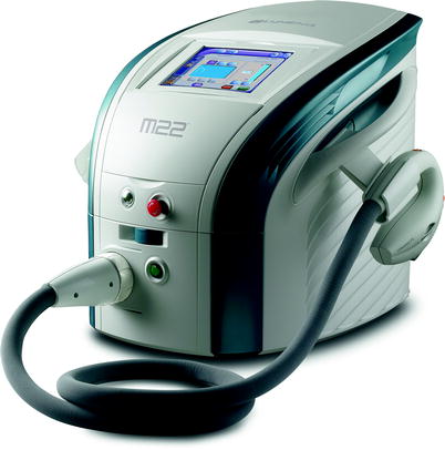
Fig. 3
(a) Patient with port-wine stain before treatment. (b) Patient after port-wine stain after treatment with IPL
Fig. 4
M 22, a new generation pulsed light device
In a certain multi-center study, it was shown that PhotoDerm VL yielded good results in both therapy resistant and hypertrophic PWS (Table 2).4
Table 2
Suggested parameters for photorejuvenation for various commercially available systems
Device | Filters (nm) | Fluence (J/cm2) | Delay time (ms) | First pulse (ms) | Second pulse (ms) | Number of sequential pulses |
|---|---|---|---|---|---|---|
Lumenis M 22 (Fig. 4) | 560 | 20–35 | 5–150 | 4–10 | 4–10 | 1, 2 or 3 |
Lumenis One/Quantum | 560 | 22–28 | 5–150 | 2.4–3.0 | 3.0–4.0 | 1, 2 or 3 |
Cutera (Limelight) | 560–1,100 | 16–26 | 12–30 | N/A | N/A | N/A |
Setting B | ||||||
Starlux | Lux G | 30–40 | 10–100 | N/A | N/A | N/A |
500–670, 870–1,200 |
Pigmented Lesions
Pigmented lesions have a spectrum ranging from epidermal lentigines and café-au-lait macules to acquired Ota’s/Ito’s nevus, Becker’s nevus and congenital nevi to post-inflammatory hyperpigmentation, melasma and decorative and traumatic tattoos. IPL systems have pulse durations in millisecond range, they do not appear to be optimally suited for treating pigmented lesions still have been reports of successful use and Q-switched laser system remains the method of choice for treating benign pigmented lesion. In a study by Bjerring et al. on 96 patients with solar lentigines and melanocytic nevi were treated.50 The IPL device used for treatment was characterized by the fact that the emitted light is conditioned by two types of optical filters: a hot mirror filter and a water chamber which work together to create the desired spectral range of light. A single session of this treatment led to a 96% reduction in pigment, 74.2% clearance for lentigo solaris and 66.3% for melanocytic nevi.
In another study, treatment of solar lentigines and ephelides was performed in three to five sessions. A total of 48% of patients showed 50% improvement in pigmentation.51
With the objective of studying the efficacy and safety of a new IPL device in the treatment of melasma in Chinese patients, Li et al. treated 89 women with melasma with a total of four IPL treatments at 3-week intervals.52 Among all the results obtained, noticeable was that 77.5% of the patients obtained 51–100% improvement. Patients with epidermal-type melasma responded better to treatment than the mixed type. The patients younger than 35 years or older than 45 years responded significantly better to the treatment, especially after one session. Minimal adverse effects were observed.
Non-ablative Photorejuvenation
IPL is very commonly used for changes of photoaging as IPL has the potential to treat multiple components simultaneously including pigmentation, telangiectasias and degraded collagen. The quantitative effects of sun exposure with resultant UV damage of structural components such as collagen and elastic fibers impact the overall appearance of aging skin. Besides, genetic factors, intrinsic factors, disease processes (rosacea) and overall loss of cutaneous elasticity associated with age also have a role to play. Photorejuvenation “photofacial” has been described a dynamic non-ablative process involving the use of the IPL to reduce mottled pigmentation, telangiectasias and smoothen the textural surface of skin.2 The treatment is in the form of three to six procedures in 3–4 week intervals where the entire face is treated and the patient may return to all activities immediately (Fig. 5).
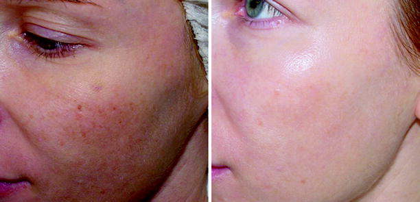
Fig. 5
Before and after pictures of a patient after 3 treatments with IPL for photorejuvenation
In a study, Zelickson demonstrated that IPL treatment results in an 81% increase in collagentype-1 transcripts while PDL treatment results in a 23% increase in Collagen type-1 transcripts improving the fine wrinkles.53 ’ 54


Related posts:


Stay updated, free articles. Join our Telegram channel
Full access? Get Clinical Tree
