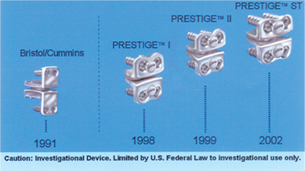
8 The Prestige family of disks traces its lineage back to the Cummins (Bristol) Artificial Cervical Joint (Fig. 8–1). The late Brian Cummins, senior consultant in neurosurgery at Frenchay Hospital in Bristol, U.K., became concerned about the need for repeated surgical therapy in patients with degenerative spinal disk disease undergoing anterior cervical fusion, particularly after two or more previous surgically or congenitally acquired fusions. Prof. Cummins recognized the potential clinical benefit of an artificial cervical joint and in 1989 began consultation with Colin Walker, head of the Department of Medical Engineering at Frenchay Hospital (DMEFH). Several concepts were considered; however, they ultimately elected to place a prosthetic joint in the intervertebral space after a standard anterior diskectomy. Several prototypes were manufactured in the DMEFH and in early 1991 the initial design was produced for clinical trial. The prosthetic joints were individually produced in the medical engineering shop and were made of Type 316 stainless steel (composition D: BS7262: part 1:1990 and ISO5832-1:1987). The prosthesis was basically a ball and socket joint that allowed flexion-extension, axial rotation, lateral bending, and, by making the ball slightly smaller than the saucer, slight translation. Fixation was initially achieved with a single anterior bone screw through each anterior flange. After the joint was placed in the first five patients, an additional screw site was added to the anterior flange of the joint and the screw sites were adjusted to ensure a more stable interface. The first five joints had simple stainless steel fracture screws. Subsequently, A-O locking screws were used to affix the joint anteriorly to the vertebral body. However, these screws were made of titanium, which added a variant metal to the process. Between February 1991 and August 1996, 22 joints were placed in 20 patients. All patients provided detailed informed consent before undergoing this innovative surgical procedure. Nineteen of the 20 cases were associated with previous surgical or congenital fusions at single and multiple levels of the cervical spine. Patients were operated because of symptomatic adjacent segment cervical stenosis, spondylosis, or herniated disk with associated myelopathy, radiculopathy, or, in one case, intractable pain. The joint appeared to be stable, it preserved motion, and it was both biomechanically and biochemically compatible. Subsidence was not observed despite several incidences of fixation screw fracture. After review in July 1996 of the medical records, examination of the patients, and obtaining delayed cervical spine motion x-rays, the results revealed this prosthesis to be suitable for cervical disk replacement.1 Figure 8–1 Metal/metal cervical disk clinical history.
Prestige Cervical Artificial Disk
 Prestige LP Surgical Technique
The Prestige I
Stay updated, free articles. Join our Telegram channel

Full access? Get Clinical Tree


 The Prestige I
 The Prestige II Disk
 The Prestige ST
 The Prestige STLP Disk
 The Prestige LP
 Conclusion


