Fig. 11.1
Model of proposed sequence of events leading to necrosis of tissue. Deformation of tissue leads to damage when the threshold is met (T def 1) and cells will start a programmed cell death leading to necrosis. If the deformation of the cell exceeds its tolerance, the cells will die immediately from necrosis. Cells can also be injured from ischemia, reducing the metabolic substrates needed and leading to anaerobic metabolism and accumulation of lactic acid. Both cellular starvation and acidification lead to apoptosis and cellular necrosis (From Stekelenburg [16])
Restoration of blood flow to an ischemic area of tissue, or reperfusion, has recently been suggested as a cause of more damage to the injured area, causing a pressure ulcer to enlarge or fail to heal. One mechanism of harm to tissue may be from reduction in capillary density from repeated loading and unloading along with ischemic insult [10]. The time to develop necrosis is reduced in patients with impaired circulation, such as those with peripheral vascular disease or hypotension.
Shear is also a cause of pressure ulcers and undermining in existing ulcers. Shear is a tangential (angular) force associated with movement, for example, sliding down in bed or being pulled over to the side of the bed. Shear forces distort blood vessels in the skin, making the effect of pressure more deleterious because the tissue is already hypoxic. Research has shown that positioning a patient at 45° head of bed elevation leads to the most detrimental combination of pressure and shear on the sacrum, because the shear stresses combined with pressure cause greater obstruction and distortion of capillaries in skeletal muscle around bony prominences than does pressure alone [11].
Microclimate, the moisture and heat of the skin, increases the risk for pressure ulcers because the moisture macerates the skin. The boggy skin does not glide against bed sheets and leads to superficial tissue injury. Skin exposure to urine or stool also injures the skin and increases risk of tissue damage from pressure and shear. Gefen [12] has provided a recent theoretical explanation of the ways in which a changing microclimate may influence the development of superficial pressure ulcers. From a mathematical model, Gefen postulated that four microclimate changes may influence pressure ulcer development – increases in skin temperature, increasing ambient temperature, increasing relative humidity, and decreasing the permeability of sheets or clothing.
11.3 Differential Diagnosis
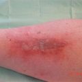
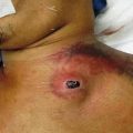
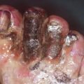
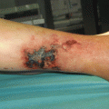
Pressure necrosis appears on tissue that has been subjected to intense pressure in patients who cannot feel the pressure or respond to it and change positions. High-risk patients and the common locations for pressure necrosis are as follows:
Position | Common location of pressure necrosis | Example pressure necrosis leading to deep tissue injury |
|---|---|---|
Flat in supine position (e.g., during surgery, hypotensive) | Buttocks tissue, unless patient is quite thin, with no buttocks tissue | 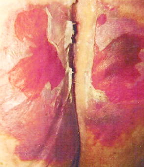 |
Necrosis appears bilaterally | ||
Supine with head of bed elevated 30–45° or slouching in a chair | Sacrum and adjacent buttocks tissue | 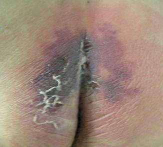 |
Sitting erect in chair | Ischial tuberosities | 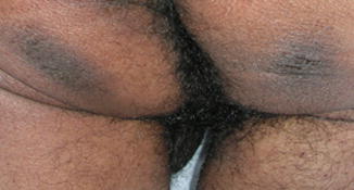 |
Supine with heels on the bed | Posterior heel in patients with immobile legs, neuropathic legs, or peripheral vascular disease | 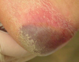 |
Wearing medical devices that are tight | Bridge of the nose from noninvasive positive pressure masks, behind the ears from oxygen tubing, shin, top of foot, and along Achilles tendon from stockings | 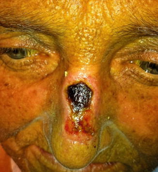 |
Differential Diagnosis
Abscess
Bruising
Cellulitis
Critical limb ischemia
Fournier’s gangrene
Hematoma/Morel–Lavellée lesions
Incontinence-associated dermatitisRelated posts:

Stay updated, free articles. Join our Telegram channel

Full access? Get Clinical Tree