Practical clinic procedures
Dermatologists make use of several diagnostic and therapeutic procedures in their everyday clinical practice.
Diagnostic procedures
The ability to diagnose a skin disease is improved by the use of better methods of observing lesions and by appropriate use of samples for laboratory investigation. Patch tests and prick tests are described on page 126.
Dermoscopy
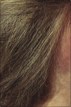
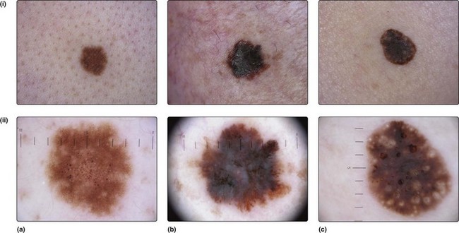
A hand lens helps when looking at small lesions such as nits on hair shafts (Fig. 1) or scabetic burrows, but dermoscopy gives added information, especially for pigmented lesions. Dermoscopy employs a ×10 magnification illuminated lens system, which can visualize a lesion after the application of a drop of oil or water between the skin and the applied lens. Detailed visualization of the epidermal structures is possible, particularly the pigment network (Fig. 2). Analysis takes account of:
Dermoscopy allows an opinion to be made about the nature and malignant potential of the lesion.
Microbiology samples
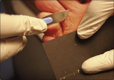
 The active scaly edge of an eruption is sampled using a disposable scalpel blade held vertically to the skin.
 Nail samples are taken from the distal portion or from debris beneath the nail using clippers or a scalpel.
 Hair sampling requires plucking of hairs as the hair root is often infected (a scalp scraping is also worthwhile).
Samples are taken onto a small sheet of black paper or a microscope slide (Fig. 3). Direct microscopy of scrapings mounted in 20% potassium hydroxide solution will show hyphae (Fig. 4).
Stay updated, free articles. Join our Telegram channel

Full access? Get Clinical Tree


