(1)
Department of Neurosurgery Faculty of Medicine, Saga University, Saga, Japan
Keywords
Transpetrosal approachAnterior transpetrosal approachPosterior transpetrosal approachMastoidectomySuperior petrosal sinus19.1 Introduction
The transpetrosal approaches (TPAs) are skull base approaches through the petrous bone. Using these approaches, the upper cerebellopontine angle (CPA) and space anterior to the upper brainstem can be exposed without strong retraction of the cerebellar hemisphere after removing the petrous bone. TPAs are broadly divided into anterior and posterior TPAs based on their relationships to the arcuate eminence [6, 21]. Both TPAs finally expose the upper CPA, but their route, procedures, and surgical field exposure differ from each other. Recently, a combined “modern” TPA, which is a combination of the anterior and posterior approaches, has also been utilized [3, 5, 6, 16]. We will describe these two TPAs separately, because an understanding of the two standard TPAs and their differences is essential.
The posterior TPA, in which the petrous bone is removed posterior to the arcuate eminence, affords a wide exposure, minimizing operative distance and brain retraction [1, 7, 8, 11, 28, 37, 39, 40, 43, 50]. These advantages are only fully realized by division of the superior petrosal sinus and tentorium and mobilization of a completely skeletonized sigmoid sinus, first described by Hakuba A and colleagues [10, 11]. Subsequently, Al-Mefty O, Spetzler RF, Ohata K, and colleagues have developed and popularized the approach [1, 6, 8, 28, 30, 39, 45]. In addition to the standard posterior TPA, Gross BA et al. reported the following three modified approaches [7]:
Extended posterior TPA
Combined modern TPA
Preservation of the superior petrosal sinus
On the other hand, in the anterior TPA, the petrous bone anterior to the arcuate eminence—Kawase’s triangle—is removed, and lesions are approached after dividing the anteromedial part of the superior petrosal sinus passing epidurally through the middle cranial fossa [19–25, 27, 44, 51]. The anterior TPA was originally described by Bochenek Z and Kukwa A in 1975 [2]. Popularized by Kawase T et al., it is sometimes referred to as the Kawase approach. Kawase T et al. first reported this approach for aneurysms of the lower basilar artery and developed it for petroclival meningiomas [20–27].
19.2 Posterior Transpetrosal Approach and Mastoidectomy
The posterior TPA is anatomically logical and comprehensible. First, it is necessary to study mastoidectomy [12, 38]. Mastoidectomy is further subdivided, based on the extent of resection of the petrous bone, into three approaches [38]:
The retrolabyrinthine approach
The translabyrinthine approach
The transcochlear approach
Furthermore, to perform the posterior TPA, it is necessary to master the technique of removing the bone over the sigmoid sinus to expose the sinus from the origin of the transverse sinus to the jugular bulb. It is also necessary to learn how to protect the vein of Labbé, which is one of several obstacles to this approach [41]. First, we will explain the surgical anatomy and step-by-step procedures of mastoidectomy [36]. Once mastoidectomy is mastered, the infralabyrinthine approach combined with the lateral suboccipital approach can be performed safely for jugular foramen lesions [34]. The infralabyrinthine approach is a variant of this approach, which will be described in detail in Chap. 20 “Microsurgical Anatomy of and Surgical Approaches to the Jugular Foramen.”
19.2.1 Microsurgical Anatomy for Mastoidectomy
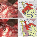
Landmarks on the skull surface include the zygomatic arch, temporal line, Henle’s spine (the suprameatal spine), and the incisura mastoidea; the most important of them are the temporal line and Henle’s spine (Fig. 19.1a) because the temporal line indicates the bottom of the middle cranial fossa and Henle’s spine indicates the underlying mastoid antrum. While drilling the mastoid air cells in the temporal bone, it is essential to confirm the sigmoid sinus, dural plate of the middle cranial fossa, mastoid antrum, compact bone, and fossa incudis (Fig. 19.1b). The triangular bone surrounded by the sigmoid sinus and dural plate of the middle cranial fossa is termed Trautmann’s triangle, which represents the biggest obstacle to approaching the intracranial region. The prominence of the compact bone is composed of the three semicircular canals (posterior, lateral, and superior). The compact bone containing the lateral semicircular canal protrudes as a prominence toward the mastoid antrum (Fig. 19.1b). The inferior edge of the prominence is the fossa incudis, where cranial nerve (CN) VII, running from the middle cranial fossa, changes its direction to descend around the external auditory canal toward the stylomastoid foramen.
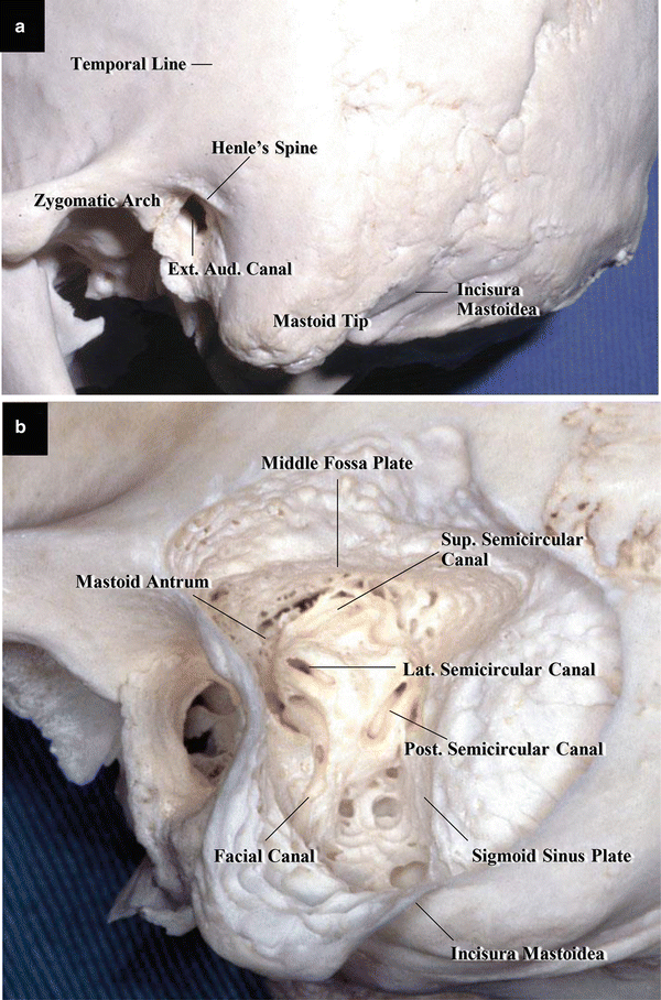
Fig. 19.1
Basic anatomy of the mastoid bone for mastoidectomy. Dry specimen, left side. (a) External view of the left temporal bone: The temporal line indicates the base of the middle cranial fossa, and Henle’s spine indicates the mastoid antrum (from Matsushima T [33] with permission). (b) External view of the left temporal bone following mastoidectomy (from Hitotsumatsu T et al. [12] with permission). The shape of mastoidectomy is triangular. The upper margin is the dural plate of the middle cranial fossa, the anterior margin is CN VII in the facial canal, and the posterior margin is the sigmoid sinus
19.2.2 Procedures of Mastoidectomy
When mastoidectomy is combined with craniotomy, we recommend first exposing the dura mater in the middle and posterior cranial fossae and posterior margin of the sigmoid sinus before starting mastoidectomy to facilitate good orientation (Fig. 19.2a–c). There are two ways to start: One is to start drilling the bony cortex at Henle’s spine and the other is to start performing osteoplastic mastoidectomy. Figure 19.2b shows the starting point for drilling the bony cortex at Henle’s spine. The procedure of osteoplastic mastoidectomy will now be explained.
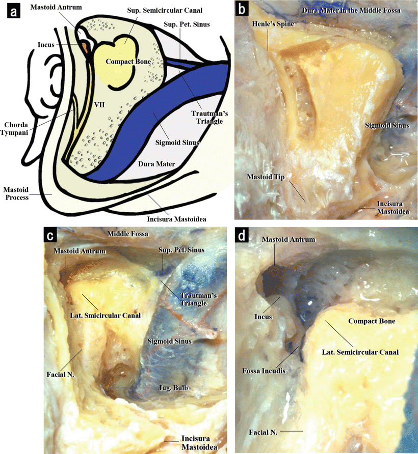
Fig. 19.2
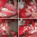
Mastoidectomy, left side. (a) Illustration of mastoidectomy on the left side. The mastoid antrum, compact bone, and cranial nerve (CN) VII have been exposed (from Matsushima T [33] with permission). (b) External view of the left mastoid bone (wet specimen). Temporosuboccipital craniotomy and lateral suboccipital retrosigmoid craniotomy have been performed, and the sigmoid sinus has partially been exposed. At the start of mastoidectomy, the bone near Henle’s spine should be drilled (from Matsushima T [33] with permission). (c) Low-magnification image after mastoidectomy (from Hitotsumatsu T et al. [12] with permission): After confirming the mastoid antrum, the compact bone is entirely exposed. Trautmann’s triangle is also exposed. Near the digastric groove, the sigmoid sinus changes its course and gets close to CN VII. (d) High-magnification image of the region around the mastoid antrum (from Hitotsumatsu T et al. [12] with permission): The lateral semicircular canal protrudes into the mastoid antrum. The incus can be seen in the antrum, and CN VII courses in the fossa incudis
19.2.2.1 Osteoplastic Mastoidectomy
During osteoplastic mastoidectomy, about 7–8 mm of the surface of the mastoid bone is removed when a bone flap of the osteoplasty is made; this not only achieves osteoplasty but also simplifies drilling the mastoid air cells. In patients with well-developed mastoid cells, the mastoid antrum can be identified only after removal of the bone flap of the osteoplasty.
The first step toward successful mastoidectomy is to detect the mastoid antrum at an early stage (Fig. 19.2d). When drilling inside the mastoid air cells, care should be taken to avoid damaging the semicircular canals or CN VII because identifying structures inside the mastoid bone is extremely difficult.
19.2.2.2 Exposure of the Dural Plate of the Middle Cranial Fossa and Upper Part of the Sigmoid Sinus
While we remove the mastoid air cells, we had better drilled the petrous bone in a triangle shape bordered by the dural plate of the middle cranial fossa, sigmoid sinus, and margin of the external auditory canal (Fig. 19.2a). First, the upper part of the petrous bone near the dural plate of the middle cranial fossa and Trautmann’s triangle should be drilled and removed, deepening the anterosuperior part to facilitate finding the mastoid antrum (Fig. 19.2b). The procedures and techniques used to expose the sigmoid sinus include:
Thinning the bone over the sinus until the sinus is visible through it
Inserting cotton into the aperture between the covering bone and dura mater of the sigmoid sinus
Removing the thin bone over the sinus using Kerisson’s punches
The insertion of cotton into the aperture between the bone and dura mater over the sinus is one method of stripping the dura mater. The sigmoid sinus is followed and exposed from above downward.
19.2.2.3 Confirmation of the Mastoid Antrum and Exposure of the Compact Bone
The mastoid antrum should be confirmed at an early stage. After confirmation of the bony prominence containing the lateral semicircular canal, protruding into the mastoid antrum, the air cells and compact bone in the shape of the three semicircular canals must be drilled, taking care not to injure the semicircular canals (Fig. 19.2c, d). To expose the mastoid antrum, it is necessary to start drilling from the area superior to rather than posterior to the porus acusticus externus. Because CN VII runs in the deep area of the upper part of the petrous bone, its injury is unlikely (Fig. 19.2c). The upper part of the mastoid bone is drilled close to the wall of the external acoustic meatus. When the shape of the compact bone becomes sufficiently clear to anticipate the course of CN VII, the first step of mastoidectomy is complete.
19.2.2.4 Complete Removal of Bone in Trautmann’s Triangle
Next, it is necessary to drill completely the posterosuperior part of the mastoid bone known as Trautmann’s triangle because it is the biggest obstacle when the dura mater is reflected. During this step, the dural plates of the middle cranial fossa and presigmoid posterior cranial fossa are exposed, and the superior petrosal sinus can be seen to run in the middle of both dural plates (Fig. 19.2c). Moreover, mastoidectomy must be extended forward to expose the dural plate of the middle cranial fossa toward the superior semicircular canal and dural plate of the posterior cranial fossa toward the jugular bulb following the sigmoid sinus inferiorly. During the exposure of the dural plate of the posterior cranial fossa, the fold of the dura mater of the vestibular aqueduct will be encountered.
19.2.2.5 Confirmation of the Facial Nerve at the Fossa Incudis and Its Downward Course
Next, the fossa incudis must be identified to confirm CN VII. The bone adjacent to the inferolateral end of the lateral semicircular canal is carefully drilled to find the fossa incudis, where CN VII runs (Fig. 19.2d). The nerve, which originates from the middle cranial fossa, changes direction at the fossa incudis to descend toward the stylomastoid foramen. It is visible through the translucent thinned bone. When the bone covering CN VII is drilled and CN VII is followed inferiorly, it is possible to see the nerve running downward into the stylomastoid foramen (Fig. 19.2c).
19.2.2.6 Exposure of the Inferior Part of the Sigmoid Sinus and Jugular Bulb
After confirming the course of CN VII, the inferior part of the mastoid bone is removed, further exposing the inferior part of the sigmoid sinus. When the sigmoid sinus abruptly changes course anteriorly, care must be taken not to injure CN VII running toward to the stylomastoid foramen in the shallow area superoanterior to the sinus (Fig. 19.2a, c). This is because the nerve gets close to the sinus in the lower part of the petrous bone. At this level, the sinus and CN VII course at two different depths, but they get close to each other and sometimes cross in the surgeon’s line of sight through the operative microscope.
19.3 Posterior Transpetrosal Approach
19.3.1 Procedures of the Posterior Transpetrosal Approach
The posterior TPA enables operating on lesions in the upper CPA from Meckel’s cave to the internal auditory canal (IAC), especially in the petroclival region, without strong retraction of the cerebellar hemisphere. The approach is utilized for the surgeries of trigeminal neurinomas, petroclival meningiomas, tumors ventral to the pons, or basilar trunk aneurysms [1, 11, 28, 29, 37, 40, 43, 45].
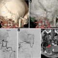
Hearing in both ears should be examined preoperatively; it is also necessary to check the size of the sigmoid sulcus, development of the mastoid air cells, mastoid antrum, and compact bone using preoperative bone computed tomography (CT). Contrast-enhanced 3D CT or cerebral angiography is also essential: the flow and size of the sigmoid sinus on both sides, flow of the superior petrosal sinus, and draining points and sizes of the superior petrosal veins (Sup. Pet. Vs.) and vein of Labbé must be checked on the operative side. Regarding intraoperative monitoring, a CN VII stimulator and/or auditory brainstem response testing should be used, depending on the case.
19.3.1.1 Extraction of Adipose Tissue, Lumbar Drainage, and Positioning
Before positioning, abdominal subcutaneous fat is extracted to fill the bony defect during closure of the scalp. Lumbar drainage is also prepared. Then, the patient is placed in a lateral recumbent position, and his/her chin is tilted slightly anteriorly. In this approach, we operate on patients by switching from the subtemporal to the lateral suboccipital retrosigmoid approaches. Therefore, the operating table and surgical field should be set up so that we can move around the patient’s head from the parietal to suboccipital regions.
19.3.1.2 Skin Incision, Muscle and Periosteal Flaps, and Craniotomy
The skin incision and temporosuboccipital craniotomy are performed as shown in Fig. 19.3a, b. Because the craniotomy is made above and below the transverse sinus, the skin incision is performed around the auricle. One or two skin flaps can be made. Because we usually make two skin flaps, a skin incision with two skin flaps is illustrated (Fig. 19.3a, b). Care should be taken not to make the skin incision smaller than the planned size, especially frontward on the temporal side. The skin is incised to avoid any inadvertent incision into the muscle and fascia, so that the temporal muscle, periosteum, and sternocleidomastoid muscle remain intact on the scalp. After making and reflecting the skin flaps, the muscle and periosteal flaps are made with a stem, which will be used during dural closure.
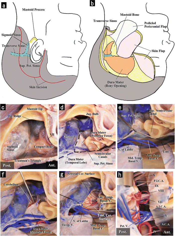
Fig. 19.3
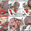
Posterior transpetrosal approach, left side. (a) Illustration of the skin incision on the left side. The transverse, sigmoid, and superior petrosal sinuses are indicated by the dashed line. (b) Illustration of the bony opening on the left side. The craniotomy extends from the supratentorial area to the infratentorial area. (c) Mastoidectomy on the left side in a cadaveric specimen in the lateral position. The dura mater in the posterior and middle cranial fossae and the compact bone have been exposed after mastoidectomy. (d) Dural incision. Two dashed lines on the exposed dural mater indicate the dural incision. The dura mater in the middle cranial fossa is incised parallel to the superior petrosal sinus, and that in the posterior fossa is incised parallel to the sigmoid sinus. The latter crosses the superior petrosal sinus. (e) Incision of the supratentorial dura mater and exposure of the cerebellar tentorium. After opening the supratentorial dura mater, the left temporal lobe is lifted, and the cerebellar tentorium can be seen. The vein of Labbé and two temporal bridging veins can also be seen in the posterior part of the dural opening and form obstacles. (f) Incision of the presigmoid dura mater. The presigmoid dura mater in the posterior fossa has been incised parallel to the sinus. Next, the posterior part of the superior petrosal sinus must be ligated and sectioned. (g) Sectioning of the superior petrosal sinus and tentorium cerebelli. The superior petrosal sinus has been incised. The tentorium has also been incised to the tentorial edge. The petrosal cerebellar surface is visible. (h) Observation of the deep area. The superior petrosal vein and cranial nerves (CNs) V, VII, VIII, and the lower CNs are visible. The space between CN V and CNs VII and VIII exists in the center of the operative field. The superior cerebellar and anterior inferior cerebellar arteries are also visible
Before mastoidectomy, the craniotomy is made above and below the transverse sinus with either one or two bone flaps. When the craniotomies are complete, the mastoid bone appears within the triangle shape (Fig. 19.3b). The anatomy and relations of temporal bone become clear, and mastoidectomy becomes easier.
19.3.1.3 Mastoidectomy via the Osteoplastic Method
When mastoidectomy is performed via the osteoplastic method, first the posterior margin of the sigmoid sinus is partially exposed through a combined craniotomy, and then the cortex of the mastoid bone is cut with a bone saw to avoid injuring the sigmoid sinus. When the bone flap of the cortex is removed, the mastoid air cells are exposed. The exposed bone is drilled and removed from the upper area. The dural plate of the middle cranial fossa and upper part of the sigmoid sinus are exposed. Here the mastoid antrum and the round lateral bulge into its cavity containing the lateral semicircular canal can be confirmed in the early stage. After the compact bone of the lateral semicircular canal is exposed, the fossa incudis, where CN VII changes course, can be identified (Fig. 19.3c). However, it is unnecessary to expose the nerve itself, and knowing its location suffices. Trautmann’s triangle is removed as described in Sect. 19.2.2 “Procedures of Mastoidectomy.”
To expose the sigmoid sinus, the bone covering the sinus is first drilled until thin, and then the thinned bone is separated from the sigmoid sinus by inserting a sheet of cotton and chiseling with a rongeur. Regarding intraoperative observation, Goto T and Ohata K stated that tight adhesion of the sigmoid sinus to the bone was not found at any part except for region where the transverse sinus joins the sigmoid sinus [6].
Related posts:
 The Veins of the Posterior Cranial Fossa: Nomenclature
 The Cerebellopontine Angle: Basic Structures and the “Rules of Three”
 Microvascular Decompression Surgery for Hemifacial Spasm: The Lateral Suboccipital Infrafloccular Approach
 Occipital Artery–Posterior Inferior Cerebellar Artery Bypass Surgery
 Microvascular Decompression for Glossopharyngeal Neuralgia: Surgical Approaches Depending on the Offending Artery
 Microsurgical Anatomy of the Cerebellomedullary Fissure and Variations of the Transcerebellomedullary Fissure Approach
Stay updated, free articles. Join our Telegram channel

Full access? Get Clinical Tree


