Chemokines
CXCL1 (growth-related oncogene-α)
CXCL4 (PF-4)
CXCL5 (epithelial neutrophil-activating peptide-78)
CXCL7 (β-TG)
CXCL8 (interleukin-8)
CXCL12 (stromal cell-derived factor-1)
CCL1 (I-309)
CCL2 (macrophage chemotactic protein-1)
CCL3 (macrophage inflammatory protein-1α)
CCL5 (RANTES)
CCL7 (macrophage chemotactic protein-3)
CCL17 (thymus and activation-regulated chemokine)
Growth Factors
Platelet-derived growth factor
TGF-β
Vascular endothelial growth factor
Insulin-like growth factor
Cytokines
Interleukin-1β
Lipid Mediators
Platelet-activating factor
Prostaglandin Ε2
Microparticles
Antibacterial proteins
Thrombocidins 1 and 2
Adhesion Molecules
P-selectin
CD40 ligand
Integrins (α2β1, α5β1, α6β1, αLβ2, αIIbβ3, αvβ3)
Thrombospondin
Fibrinogen
Fibronectin
Others
Albumin
IgG
IgM
IgA
Table 14.2
Dense granule contents
Nucleotides |
Adenosine disphosphate |
Adenosine triphosphate |
Ions |
Ca |
Mg |
P |
Pyrophosphate |
Serotonin |
Epinephrine |
Histamine |
14.2.2 Surface Molecules Expressed on Platelets
14.2.2.1 P-Selectin
P-selectin (CD62P) is an integral membrane glycoprotein that is located in membranes of platelet α-granules [54]. Upon platelet activation, P-selectin is rapidly expressed on the cell surface and secreted into plasma [28, 45]. There are approximately 1,000 P-selectin molecules in an unstimulated platelet and 10,000 in an activated platelet [46]. By binding its major ligand, P-selectin glycoprotein-1 (PSGL-1), which is expressed on almost all leukocytes, platelet P-selectin supports adhesion of polymorphonuclear cells [36], monocytes/macrophages [17], and lymphocytes [38].
14.2.2.2 CD40 Ligand
CD40 ligand (CD154) is a member of the tumor necrosis factor (TNF) superfamily and is present in platelet α-granules. Platelets can interact with a large number of CD40-bearing immune cells such as B cells, macrophages, and dendritic cells, and nonimmune cells such as endothelial cells [49]. Platelet CD40 ligand interacts with and stimulates endothelial cells through the CD40 receptor, and this results in increased expression of adhesion molecules and chemokines (Fig. 14.1) [23]. In addition, ligation of CD40 by platelet-derived CD40 ligand induces dendritic cell maturation and B-cell isotype switching, and increases CD8+ T-cell responses [13].
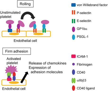
Fig. 14.1
Interaction of platelets with the endothelium via adhesion molecules
14.2.2.3 Integrins
Integrins are a family of adhesion molecules present on most cell types that mediate cell–cell and cell–matrix interactions. Platelets have six different integrins (α2β1, α5β1, α6β1, αLβ2, αIIbβ3, and αvβ3) in α-granules [9]. On platelets, αIIbβ3 (glycoprotein (GP) IIb/IIIa) is the major integral plasma membrane protein. αIIbβ3 is the only integrin expressed uniquely on platelets and mediates firm adhesion of platelets to the intercellular adhesion molecule (ICAM)-1 on the endothelium (Fig. 14.1) [8].
14.2.2.4 Chemokine Receptors
Platelets express many kinds of chemokine receptors , CCR (CCR1, CCR2, CCR3, CCR4, CCR5, CCD6, CCR7, CCR8, CCR9, and CCR10) and CXCR (CXCR1, CXCR 2, CXCR 3, CXCR4, and CXCR5) on their surface. Platelet receptors are all members of the G protein-coupled receptor family, similarly to those on other cell types. Several chemokines exert major effects on platelet functions of aggregation and adhesion [18], and platelet chemokines themselves can directly activate platelet function.
14.2.2.5 Toll-Like Receptors (TLRs)
TLRs are a family of pattern recognition receptors that are expressed by immune cells such as macrophages, dendritic cells, granulocytes, natural killer cells, and T cells, and by nonimmune cells such as fibroblasts and epithelial cells [27, 33]. Expression of TLR1-9 on platelets has been detected [51] and platelets can recognize pathogens via TLRs. Platelet-expressed TLRs can modulate sepsis-induced thrombocytopenia and TNF production in vivo [3].
14.2.2.6 High-Affinity Receptor for IgE (FcεRI)
FcεRI is expressed on the surface of mast cells, basophils, dendritic cells, and monocytes, and plays a key role in immediate hypersensitivity and IgE-mediated delayed-type hypersensitivity reactions. Platelets also express FcεRI on their surface, in addition to FcεRII and FcεRIII [21, 29]. Engagement of FcεRI on platelets induces release of serotonin (5-hydroxytryptamine, 5-HT) and regulated on activation, normal T cell expressed and secreted (RANTES, CCL5).
14.2.3 Soluble Mediators in Platelets
14.2.3.1 Chemokines
Chemokines are small chemoattractant proteins that stimulate migration and activation of cells, especially phagocytic cells and lymphocytes, and have a central role in inflammatory responses. The first platelet chemokine, platelet factor 4 (PF-4, CXCL4), was described in the 1960s [14]. A number of chemokines found in platelet α-granules are rapidly secreted upon platelet activation [18]. CXCL4 and β-thromboglobulins (β-TG, CXCL7) are particularly abundant in platelets.
14.2.3.2 Platelet-Derived Microparticles (PDMPs)
During platelet activation, microparticles are formed from the surface membrane of platelets by an exocytotic budding process, and are released into the extracellular space [24]. These PDMPs range in size from 0.02 to 0.1 μm and have both pro- and anticoagulant activities [60]. The microparticles bind to leukocytes, endothelial cells, and subendothelial matrix [47], and subsequently enhance adhesion of monocytes [5] and granulocytes [15].
14.2.3.3 Antibacterial Proteins
Antibacterial proteins are important components of the innate immune system that are found in many organisms and produced by various types of cells. These proteins bind to and disrupt bacterial membranes. Platelets store antibacterial proteins in α-granules and the proteins are released after cell activation [34]. These molecules are referred to as thrombocidins. Thrombocidin 1 and thrombocidin 2, which are related to the CXC-chemokine family, are lethal to various bacterial species such as Staphylococcus aureus [34].
14.2.3.4 Serotonin
Serotonin (5-HT) is a monoamine neurotransmitter that is biochemically derived from tryptophan and is stored in platelet-dense granules and mast cell granules. 5-HT affects the activation, migration, phagocytosis, cytokine production, and apoptosis of T cells, B cells, monocytes, and dendritic cells [10, 12, 25, 26, 32, 35, 52, 61].
14.3 Platelet Interactions with Leukocytes and the Endothelium
14.3.1 Interaction of Platelets with the Endothelium
Under normal physiological conditions, platelets circulating in blood do not interact with nonactivated endothelium. On activation, endothelial cells express adhesion molecules such as von Willebrand factor, P-selectin, and E-selectin that mediate platelet rolling (Fig. 14.1) [6]. Platelet–endothelial cell interactions induce full activation of platelets and surface expression of P-selectin, leading to firm adhesion of platelets on the endothelium.
14.3.2 Interaction of Platelets with Leukocytes
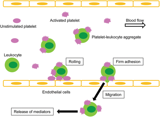
Platelets affect the function of leukocytes, including neutrophils, T cells, natural killer cells, B cells, monocytes, macrophages, and dendritic cells via direct cell–cell contacts and/or via soluble mediators (Table 14.3). Cell–cell contacts between platelets and leukocytes can occur in two ways: (1) leukocyte adhesion to activated platelets that are adherent to the endothelium or the extracellular matrix, and (2) intravascular formation of platelet–leukocyte complexes [6]. In both cases, the interaction between platelet P-selectin and leukocyte PSGL-1 is the first step of a complex formation, leading to activation of integrins on the leukocyte surface and firm adhesion of leukocytes to activated platelets (Fig. 14.2).
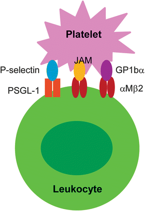
Table 14.3
Influence of platelets on the function of leukocytes
Increase | Decrease | |
|---|---|---|
Neutrophil | Adhesion | |
Migration | ||
Mediator release | ||
T helper cell | Adhesion | Proliferation |
Migration | Mediator release | |
Proliferation | ||
T cytotoxic cell | Adhesion | |
Migration | ||
Proliferation | ||
Mediator release | ||
Cytotoxic activity | ||
B cell | Adhesion | |
Migration | ||
Proliferation | ||
Isotype switching | ||
Germinal center formation | ||
Antibody production | ||
Natural killer cell | Adhesion | Cytotoxic activity |
Migration | ||
Monocyte/macrophage | Adhesion | Mediator release |
Migration | ||
Mediator release | ||
Differentiation to macrophages | ||
Proteolysis | ||
Dendritic cell | Maturation | Maturation |
Mediator release | Phagocytic activity |
Fig. 14.2
Interaction of platelets with leukocytes via adhesion molecules. JAM junctional adhesion molecule
14.4 Role of Platelets in the Pathogenesis of Inflammatory Diseases and Conditions
14.4.1 Allergic Skin Diseases
14.4.1.1 Platelet Activation in Atopic Dermatitis
Platelets are increasingly recognized as important in allergic skin inflammation. Atopic dermatitis is a recurrent pruritic eczematous skin disease (see Chap. 22). In patients with atopic dermatitis, scratching due to itch often results in excoriation and subsequent platelet aggregation at the inflamed skin lesion, implying that blood platelets may be activated compared with those in healthy individuals. Serum levels of PF-4 and β-TG, and plasma levels of PDMPs and soluble P-selectin, which are widely known as platelet activation markers, are significantly increased in patients with atopic dermatitis [30, 56–58]. Plasma levels of serotonin are also elevated in patients with atopic dermatitis [52]. Interestingly, the levels of these mediators correlate with disease severity. These findings suggest that platelets circulate in an activated state in patients with atopic dermatitis and are involved in the pathogenesis of this disease.
14.4.1.2 Role of Platelet P-Selectin in Allergic Skin Diseases
In animal models of allergic dermatitis, platelets have been shown to be important for recruitment of leukocytes into inflamed skin through formation of platelet–leukocyte aggregates via P-selectin on platelets in blood [20, 55, 57, 58]. The platelet–leukocyte aggregates are formed in circulating blood, roll along the endothelium, and transmigrate into subendothelial tissue (Fig. 14.3). Platelet P-selectin also contributes to the process of antigen sensitization [42]. These findings suggest that platelet P-selectin is important for development of cutaneous allergic reactions.