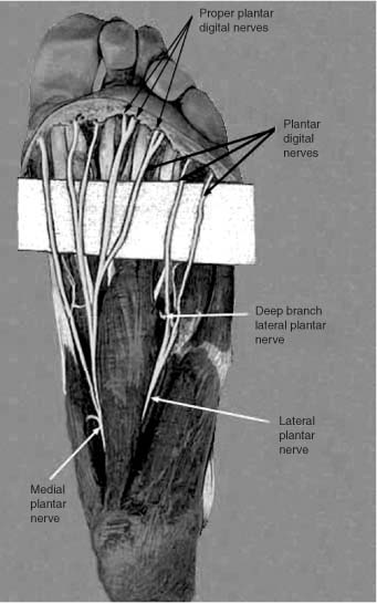
15 The tibial nerve enters into the foot behind the medial malleolus, deep to the flexor retinaculum and between the tendons of the flexor hallucis longus and the flexor digitorum longus. The nerve then divides into its terminal branches, the medial and lateral plantar nerves.1 The medial plantar nerve, homologous to the median nerve in the hand, is the larger of the two plantar nerves. The nerve runs first on the lateral side of the medial plantar vessels deep to the abductor hallucis, then between the abductor hallucis and the flexor digitorum brevis, and then finally between the flexor hallucis brevis and the flexor digitorum brevis muscles. At the level of the tarsometatarsal joints the nerve ends by dividing into a proper plantar digital nerve to the skin of the medial side of the big toe and three common plantar digital nerves. Before dividing it supplies muscular branches to the abductor hallucis, flexor digitorum brevis, and flexor hallucis brevis muscles. It also gives off cutaneous branches, which pierce the plantar aponeurosis to supply the posteromedial part of the sole of the foot as well as articular and vascular twigs to adjacent joints and vessels. The common plantar digital nerves supply muscular branches to the first and inconsistently to the second lumbricales muscles. It also gives cutaneous branches to the medial two thirds of the anterior part of the sole. The common plantar digital nerves finally divide into the proper plantar digital nerves that supply sensation to the medial interdigital spaces and the plantar surfaces of the corresponding toes. Lastly, each sends an upward curving branch that supplies the nail bed. In summary, the medial plantar nerve supplies the flexor hallucis brevis muscle, the first lumbricales of the foot, the skin of the medial three and a half toes, the abductor hallucis muscle, and the flexor digitorum brevis muscle. The lateral plantar nerve, homologous to the ulnar nerve in the hand, divides off from the tibial nerve and passes outward and forward in the sole of the foot on the medial side of the lateral plantar vessels. The nerve runs between the flexor digitorum brevis and the quadratus plantae, and then between the quadratus plantae and the abductor digiti minimi. Near the base of the fifth metatarsal bone, the lateral plantar nerve gives off branches to the quadratus plantae and abductor digiti minimi muscles and cutaneous branches to supply the lateral side of the sole; it then divides into superficial and deep branches. The superficial branch splits into proper and common plantar digital nerves. The proper plantar digital nerve supplies the skin and fascia on the plantar and lateral sides of the small toe, including the tip, branches to the flexor digiti minimi and the interossei of the fourth metatarsal space, and filaments to the fifth metatarsophalangeal and interphalangeal joints. The common plantar digital nerve divides into two proper plantar digital nerves that supply the skin and fascia of the plantar aspect, sides, and nail beds of the fourth and fifth toes. The deep branch accompanies the plantar arterial arch on the deep surface of the flexor tendons. It supplies branches to the adductor hallucis; the second, third, and fourth lumbricales; the interossei in the medial three metatarsal interspaces; and twigs to the plantar arch and adjacent joints. In summary, the lateral plantar nerve supplies the flexor digiti minimi brevis muscle, the plantar and dorsal interossei, the lateral three lumbricales of the foot, the adductor hallucis muscle, the abductor digiti minimi, and the quadratus plantae (Fig. 15-1). The plantar nerves may be exposed proximally near their origin as they divide off from the posterior tibial nerve deep to the flexor retinaculum. For this exposure the reader is directed to Chapter 13. If a distal exposure of the nerves is required, they may be approached from either a dorsal web splitting incision or a plantar intermetatarsal incision. The dorsal approach is more commonly used for a first-time resection of a Morton’s neuroma. This condition most probably represents an entrapment neuropathy of the common digital nerves between the metatarsal heads and is seen most frequently between the second and third web spaces.2 Secondary neuromas warrant exposure from the plantar direction, and although problems with wound healing are negligible, the recuperation period is longer.3
Plantar Nerves
ANATOMY
POSITIONING AND SURGICAL EXPOSURE
Plantar Nerves
Only gold members can continue reading. Log In or Register to continue


Full access? Get Clinical Tree


