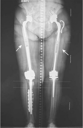
CHAPTER 57 Periprosthetic Hip Fractures
Periprosthetic hip fracture is a severe complication of total hip arthroplasty (THA). Surgical treatment of such fractures is a challenge, as it is often accompanied by fracture comminution, bone deficiency, and femoral component loosening. Of 1049 periprosthetic femoral fractures recorded in the Swedish National Register, 23% required reoperation and 18% developed postoperative complications.1 Periprosthetic hip fracture is associated with poor functional outcome, increased morbidity, and a high incidence of mortality, as well as with elevated economic burden.
Periprosthetic fracture is increasingly prevalent; it has been estimated to range from 0.1% to 3.2% for primary uncemented total hip arthroplasties and from 3% to 12% for revisions performed with or without cement. Factors associated with this increase include a broadening of THA indications, a growing population of hip joint replacement recipients, increased use of uncemented implants, and an increasing population of revision recipients. Roughly 5% of revisions performed are for periprosthetic fracture, making it the third most frequent reason for revision of primary total hip replacements. Periprosthetic fracture as a reason for revision is currently less common than either aseptic loosening or sepsis and has the same frequency as dislocation.2
RISK FACTORS
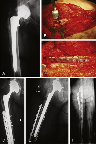
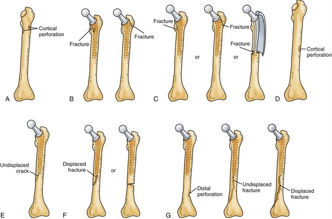
Patients at risk for intraoperative fracture include those with osteoporosis, osteopenia (as in rheumatoid arthritis), altered bone morphology or deformity (as seen in Paget disease), developmental dysplastic hip, and old proximal femoral fractures.3 Local risk factors include cementless stems (especially in revised settings), impaction bone grafting in revision procedures, minimally invasive techniques, under-reaming of the femoral cortex, use of large-diameter femoral stems, and a low ratio of femoral cortex diameter to canal diameter.4 Risk factors for postoperative periprosthetic fracture include osteoporosis, osteolysis, cortical perforation, loose femoral implants, and ipsilateral hip and knee revision arthroplasty that produces two stress risers in the isthmus of the femoral diaphysis (Fig. 57-1).5

FIGURE 57-1 An 81-year-old woman after three revisions of the right knee, primary ipsilateral total hip arthroplasty, two revisions of the left knee, and two revisions of the left hip. She underwent surgery after right knee and left hip periprosthetic fractures. She is still at high risk for recurrent periprosthetic fracture. The stress risers in both femoral diaphyses are marked with white arrows.
CLASSIFICATION OF FEMORAL FRACTURES
Many classifications of periprosthetic fractures are reported in the literature. Classifications provide information about the site of the fracture; they are also of value in determining treatment strategy. The Vancouver Intraoperative and Postoperative Classification, proposed by Duncan and Masri,6 is the most widely used system. The system is simple, reproducible, and validated, as well as being useful in formulating treatment strategies. It takes into account the site of the fracture, the stability of the implant, and the condition of surrounding bone stock: the most important factors for making the decision regarding the optimum course of treatment. The Vancouver Intraoperative Classification can be used to guide management of intraoperative fractures, although its reliability and validity in such situations have not been as widely documented.
Vancouver Intraoperative Fracture Classification
The Vancouver system takes into account fracture location, pattern, and stability, dividing fractures into three types. Type A involves the proximal metaphysis (trochanteric area); Type B is diaphyseal (tip of the stem); Type C is distal to the stem tip and not amenable to insertion of the long revision stems. Each type is further subclassified as either subtype 1 (in which there is only cortical perforation), subtype 2 (nondisplaced crack), and subtype 3, with a displaced and unstable fracture pattern (Fig. 57-2).4

FIGURE 57-2 Vancouver classification of intraoperative femoral periprosthetic fractures. A, Type A1. B, Type A2. C, Type A3. D, Type B1. E, Type B2. F, Type B3. G, Type C1 (left image), type C2 (center image), and type C3 (right image).
(From Greidanus NV, Mitchell PA, Masri BA, et al: Principles of management and results of treating the fractured femur during and after total hip arthroplasty. Instr Course Lect 52:309-322, 2003.)
Vancouver Postoperative Fracture Classification
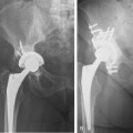
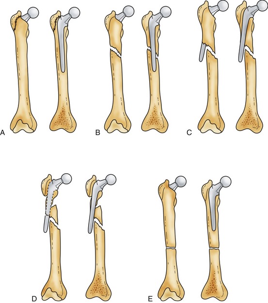
The site of the fracture, stability of the prosthesis, and quality of the bone stock determine fracture severity (Fig. 57-3). In type A, the greater or lower trochanter is affected. Type B fractures are those at or just distal to the stem tip. Type B has three subtypes: B1, in which the prosthesis is stable; B2, in which there is a loose prosthesis but adequate bone stock; and B3, in which the prosthesis is loose and there is marked proximal bone loss or damage to a degree that a standard revision component will not be supported. Type C fractures are well distal to the stem tip.

FIGURE 57-3 Illustrations demonstrating the Vancouver classification for fractures around the femoral component of a total hip arthroplasty. A, Type A fracture, which occurs at the proximal part of the femur with displacement of the greater trochanter (AGT) or lesser trochanter (ALT). B, Type B1 fracture, which occurs around or just distal to a well-fixed femoral stem. C, Type B2 fracture, which occurs around or just distal to a loose femoral stem with adequate proximal bone. D, Type B3 fracture, which occurs around or just distal to a loose stem with poor proximal bone stock. E, Type C periprosthetic fracture, which occurs well distal to the stem tip.
(From Parvizi J, Rapuri VR, Purtill JJ, et al: Treatment protocol for proximal femoral periprosthetic fractures. J Bone Joint Surg Am 86[Suppl 2]:8-16, 2004.)
MANAGEMENT OPTIONS
Femoral Intraoperative Fractures
Goals of treatment include the following:
Type A1: Metaphyseal Cortical Perforation
Type A1 fractures are usually stable and can be treated with bone graft alone—usually obtained from the acetabular reaming.
Type A2: Nondisplaced Linear Crack
Fractures of the greater trochanter should always be stabilized, even when there is no displacement and the fracture seems stable. Although there is no solid evidence for this recommendation, it is a matter of good sense to include this simple surgical step, which likely improves the general outcome. Surgical options in a trochanter fracture include placement of cerclage wire, fixation with two screws, or a hook plate fixation. Such stabilization is effective before the stem is inserted; it may prevent propagation when a proximally coated stem is used. It is important to explore the fractures of the lower trochanter to determine their extent. If a cemented stem is used, fracture should be wired beforehand to prevent cement ingress into the fracture line. If the implant is noncemented, it should also be wired in cases in which the fracture line extends below the lesser trochanter.
Type A3: Displaced or Unstable Fracture of the Proximal Femur or Greater Trochanter
Type A3 fractures are best treated with noncemented components, because cement can at the time of pressurization exit through breaches in the cortex and reduce the quality of the cement mantle. Such cement extrusion can potentially injure neurovascular structures and can also create a barrier between the fracture fragments, delaying healing. Such fractures can generally be managed using simple cerclage wires or cables if the implant provides both proximal and distal rotational control. If the implant does not provide such intrinsic stability, external stability must be obtained with strut grafts, cable plates, or claw plates. Another possible alternative is performing anatomic fracture reduction and stabilization and cementing a long stem in place.
Type B1: Diaphyseal Cortical Perforation
Diaphyseal cortical perforation is most commonly caused by incautious use of cement removal devices or reamers during canal preparation.7 If a diaphyseal cortical perforation is detected, it should be bypassed by a distance of two cortical diameters, using a longer stem. Before stem insertion, consideration should be given to cerclage fixation of the bone at or below the perforation to prevent crack propagation. If the perforation is at the tip of the longest stem, it should be bypassed with a cortical strut or plate; such a perforation should also be cancellous bone grafted at the time of strut or plate insertion and fixation (Fig. 57-4).

FIGURE 57-4 A, A 77-year-old female patient with a loose femoral stem, proximal femoral osteolysis, and liner wear. B, Cortical perforation (type B1 periprosthetic intraoperative fracture) that was produced during femoral preparation, probably secondary to the varus alignment of the previous stem. C, Cable-plate system used to bypass the cortical perforation after the implantation of a long revision stem and cancellous bone grafting. D and E, Anteroposterior and axial radiographs obtained 3 months after the revision procedure, showing the tapered fluted monoblock stem bypassing the cortical perforation, the cable-plate system stabilizing the fracture, and the acceptable integration of cancellous bone graft. F, Long leg radiograph 14 months after the revision procedure. The fracture was resolved without cortical onlay strut graft.
Type B2: Nondisplaced Linear Crack
Type B2 fractures usually result from increased hoop stress during placement of either the broach or the implant. Injuries of this type may be missed intraoperatively, only to be diagnosed on review of postoperative radiographs. If they are diagnosed intraoperatively, management can consist of placement of cerclage wire, as long as the implant is stable. If implant stability has been compromised, one should consider bypassing the defect whenever possible. Cortical allograft may be used if bone quality is deemed poor or if the fracture cannot be bypassed with the stem. A plate and screws should be considered instead, or in combination with cortical struts. If such injuries are diagnosed postoperatively, the patient may be treated with protected weight bearing for 6 weeks to 3 months, until healing has begun.
Type B3: Displaced Fracture of the Midfemur
Displaced fracture of the midfemur often occurs at the time of femoral dislocation or prosthesis reduction, through an area of weak bone. It can also occur at the time of femoral cement removal, femoral preparation, or stem implantation. When such fractures occur, they should be exposed, reduced, and fixed, either with cerclage wires or cables (in the case of oblique or spiral fractures) or with one or two cortical struts when the fracture is transverse in configuration. Once the integrity of the femoral canal is re-established, the femoral stem can be inserted; the fracture should be bypassed by at least two cortical diameters. The Wagner lateralized fluted tapered stem (Zimmer GmbH, Winterthur, Switzerland) is a good choice in treating this type of fracture. Made of Ti-Al-Nb alloy, it features a rough-blasted surface to provide axial and rotatory primary stability and good preconditions for osseointegration. Cortical allograft may occasionally be necessary.
If the fracture occurs distal to a well-impacted femoral stem and the stem is difficult to safely extract without compromising bone stock, the stem should be retained and extramedullary strut and cable augmentation should be done to stabilize the fracture fully.
Type C1: Cortical Perforation
Although rare, cortical perforation can occur during cement removal or canal preparation. If it occurs, it should be grafted. Further bypass with a cortical strut should be considered, to avoid leaving a significant stress riser that may increase the risk of postoperative fracture.
Type C2: Nondisplaced Crack Extending Just above the Knee
If a type C2 fracture is recognized intraoperatively, it should be treated with cerclage wires—with or without a cortical strut allograft—if the surgeon considers the fracture potentially unstable. If the fracture is a long, spiral, nondisplaced crack, there may be sufficient inherent stability that a strut is not required.
Type C3: Displaced Fracture of the Distal Femur That Cannot Be Bypassed with a Femoral Stem
When type C3 fractures occur, they should be treated with open reduction and extramedullary fixation such as a plate, or a plate and onlay strut cortical allograft. Unless necessary, it is our preference not to use a strut allograft, to avoid another rare but possible source of infection.

Full access? Get Clinical Tree

