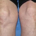
Fig. 9.1
(a) vesicles and erythematous plaques on the chest, (b) targetoid plaques on the abdomen, (c) punch biopsy of vesicle with H&E staining (From Morganroth and McHargue [4]. Reprinted with permission from American Medical Association)
There appears to be a hormonal component to disease activity, with many patients experiencing improvements in late pregnancy, but flares after delivery [5]. Flares can also occur during menstruation and with oral contraceptive use [6]. PG can present initially in the postpartum period in a reported 25 % of cases, sometimes within hours of delivery [3]. It can recur with subsequent pregnancies, sometimes earlier in the gestation and more severe in quality [3].
Pathogenesis
The histologic and immunologic features of PG are similar to those of bullous pemphigoid (BP) [7]. Like BP, PG is associated with antibodies to two hemidesmosomal proteins: BP180 (collagen XVII, a transdermal hemidesmosomal protein) and, to a lesser degree, BP230. Both antibodies are members of the IgG1 subclass [8]. The purported mechanism of PG is initiated in the placenta, where MHCII molecules are thought to be aberrantly expressed and expose the maternal immune system to the BP180 antigen. Normally, the fetal trophoblastic cells do not express these MHC molecules, so the maternal immune system is tolerant to the fetoplacental unit [9]. Because the BP180 antigen is present in both the skin and the placenta, cross-reactivity can occur, resulting in an autoimmune reaction targeting the basement membrane of the epidermis [10]. Destruction of hemidesmosomal proteins leads to vesicles and bullae. The involved autoantibodies are also capable of activating complement and precipitating infiltration of neutrophils and eosinophils [6].
Certain individuals can be predisposed to developing PG during pregnancy. There is an association between PG and HLA-DR3 in up to 80 % of patients and with HLA-DR4 in up to 53 % of patients; both are expressed in up to 50 % patients with PG, but in only 3 % of the general population [10].
Diagnosis
Diagnosis of PG can be confirmed based on clinical, histologic, and direct immunofluorescence (DIF) findings. On histology, urticarial lesions demonstrate a perivascular lymphocytic and eosinophilic infiltrate. In their plaque form, lesions demonstrate dermal edema and spongiosis as well as basal cell necrosis on dermal papillae tips. Vesiculobullous lesions demonstrate subepidermal blistering and bullae can contain eosinophils [6] (see Fig. 9.1). On DIF, lesions demonstrate linear C3 deposition at the basement membrane zone. IgG can also be present along the basement membrane [11]. DIF is the gold standard of diagnosis when combined with the appropriate clinical picture.
Indirect immunofluorescence (IIF), additionally, can detect IgG antibodies at the basement membrane in around 20 % of patients [11]. ELISA testing can also be helpful for diagnosis through detection of BP180 antibodies. In 2004, a commercially available BP180-NC16a domain enzyme-linked immunoassay became available. ELISA was shown in 2008 to have a higher sensitivity for BP and PG than IIF (93 % compared to 74 %) with a similar specificity [12].
Treatment
Remission of PG can occur without intervention within weeks of delivery [3]. In general, the pruritus associated with PG in not tolerable to patients and treatment should be started upon presentation. Immunosuppression with steroids is the mainstay of therapy, with steroid-sparing agents generally started after delivery to avoid fetal exposure to these agents.
Topical Agents/Antihistamines
Topical steroids can be started during the early stages of PG before the presence of blistering. Typically, a potent corticosteroid such as clobetasol propionate 0.05 % or betamethasone 0.05 % dipropionate can be applied twice a day [13]. They can be used along with emollients and oral antihistamines to alleviate pruritus and prevent eruption of blisters. FDA category B antihistamines are generally considered safer to use after the first trimester.
Oral Corticosteroids
Once a patient has blistering, systemic corticosteroids are typically added to the treatment plan. Oral prednisone and prednisolone are mainstays of therapy that are used during pregnancy as well as postpartum. There is some evidence for a more liberal approach to starting oral corticosteroids; a retrospective analysis of 13 patients in Iran found that those in whom oral corticosteroids were implemented sooner had faster improvement [14].
The accepted dosage for severe PG during the post-partum period is 0.5–1.0 mg/kg/day of prednisone that can be tapered when tolerated [6]. It is important to monitor patients for appropriate response and make changes as necessary. In general, a patient should respond to an appropriate dose after 3 days of treatment (as defined below); if the patient does not respond, a 2 mg/kg/day dose is given. Once the patient responds, the steroids can be tapered and maintained at the lowest effective dose [10].
The use of oral corticosteroids during pregnancy is important for the treatment of many autoimmune diseases. Along with topical agents, systemic corticosteroids are the only mainstay of therapy for PG that is used during pregnancy. Transplacental passage of steroids differs by type, with non-fluorinated corticosteroids such as prednisone largely deactivated before passage to the fetus [15]. The fluorinated compounds, meanwhile, such as betamethasone, do pass to the fetus. Prednisone is therefore the preferred corticosteroid during pregnancy.
Some studies have shown that prednisone can lead to intrauterine growth retardation, premature rupture of membranes, and preterm delivery [15]. There have also been reports of corticosteroids causing cleft palate in animals [16], with small studies replicating this finding in humans when steroids are given between 4 weeks prior to conception to 12 weeks after [17]. Larger human studies have not replicated these findings [18].
In 2013, a cohort study of over 1700 children compared survival and neurodevelopmental disability between those whose mothers were treated with one vs multiple courses of systemic corticosteroids; the study found no significant difference between the groups [19]. However, follow-up ended when children were 5 years old, leaving open the possibility of differences between groups in late-presenting neurobehavioral functioning. Chi et al., additionally, found an association with blistering diseases of pregnancy and fetuses that were born small for gestational age, but concluded that the use of oral corticosteroids was not a risk factor [20].
Once steroids are discontinued, the patient and the fetus should be examined for adrenal insufficiency, depending on the duration of use [21]. Some recommend a maximum dose of 7.5 mg/day of prednisone when use is prolonged in a pregnant patient; doses greater than 20 mg daily should be avoided [18].
Alternatives to corticosteroids can be considered in steroid-refractory cases during pregnancy, as well as postpartum to avoid the side effect profile of long-term corticosteroid use. Steroid-sparing therapies also have the potential to cause serious side effects and their risks and benefits should be considered.
Azathioprine
While there are no prospective controlled trials of azathioprine, there are several case studies showing varying degrees of benefit of this drug. It is typically given at doses of 50–150 mg/day [13]. Kreuter et al. described a patient whose disease continued to progress on 150 mg/day prednisone after 10 days of therapy. Azathioprine 100 mg/day was implemented, which improved the patient clinically and enabled prednisone to be lowered to 50 mg/day, though not below this point [22]. Cianchini et al. documented a woman with severe PG who was started on daily doses of prednisone 100 mg, azathioprine 150 mg, and dapsone 125 mg with partial response [23].
Azathioprine is a category D drug; it is therefore generally used in the postpartum period only in cases of PG [13]. However, there is data supporting its safety during pregnancy, much of which is from studies on azathioprine use for organ transplants as well as other autoimmune diseases such as systemic lupus erythematosus [18]. No associations with congenital malformations have been noted, although there may be a slightly increased risk of atrial or ventricular septal defects [24]. There have been reports of cytopenias in neonates born to women taking azathioprine, however since initiating a protocol to halve azathioprine doses at 32 weeks gestation, there have not been reports of cytopenias [25].
Dapsone
Dapsone can also be used as an adjuvant therapy for PG, typically given at 50–150 mg/day. Amato et al. reported on a patient on prednisone, azathioprine, dapsone, and plasmapheresis who had a limited response [26]. The Cianchini case described above also used both azathioprine and dapsone as adjuvant agents with limited response.
Dapsone is pregnancy category C. Prior to starting dapsone, G6PD levels should be checked to avoid hemolysis in vulnerable patients. Fetal risks include hyperbilirubinemia and hemolytic anemia; when patients use dapsone while breastfeeding, infants should be monitored for hemolysis.
Cyclophosphamide
Cyclophophamide is also category D in pregnancy and only used postpartum in severe, steroid-refractory cases of PG. One case report published in 1996 described a patient with severe persistent PG who also had anti-phospholipid syndrome. This patient achieved complete remission on cyclophosphamide 0.75 g/m2, given in monthly doses by intravenous infusion over the course of 8 weeks, and another dose 5 months later. This patient’s illness was severe enough that she was delivered by emergency c-section at 32 weeks when prednisone 120 mg/day did not control symptoms. High dose prednisone and azathioprine were unable to control the disease postpartum as well [13, 27].
Intravenous Immunoglobulin (IVIg)
Unlike most therapies for PG, IVIg is not immunosuppressive, and has a less concerning side effect profile; as such, its use has expanded in recent years [7]. Its use in pregnancy has not been shown to harm the fetus during human gestation (category C). Typically it is added to therapy when systemic corticosteroids plus adjuvant dapsone or azathioprine are unable to control blistering. As with other PG therapies, only a handful of case reports are available as evidence for the effectiveness of IVIg, all reporting a favorable response to IVIg. The dose in all was 1–2 g/kg in monthly cycles; complete remission was achieved in 3–4 months with no reported side effects [7]. As mentioned above, Kreuter et al. described a patient whose disease worsened on azathioprine and prednisone when the prednisone was tapered; upon addition of IVIg, the patient’s lesions completely resolved [22]. Rodrigues et al. reported a very similar case [28].
The use of IVIg is better established in pemphigus than PG or BP, with over 100 publications favoring the use of IVIg in pemphigus. Most of the reports utilized IVIg at a dose of 2 g/kg/cycle given over 2–5 days and showed a positive clinical outcome, decrease in pathologic autoantibodies, and a steroid-sparing effect [13].
Plasmapheresis
Plasmapheresis works by removing autoantibodies from the serum. There are a small number of case reports documenting the use of plasmapheresis in PG, both during pregnancy and after delivery. Amato et al. demonstrated a partial response when plasmapheresis was added to prednisone, azathioprine, and dapsone [26]. Van de Wiel et al. reported a patient who acquired PG in the 20th week of her pregnancy. She received plasmapheresis at 26 weeks, at delivery, and postpartum, with complete resolution of disease [29].
Immunoadsorbtion (IA)
Like plasmapheresis, IA removes autoantibodies from circulation, but can specifically remove IgG and does not require plasma product replacement. Recommendations for treatment of autoimmune blistering diseases with IA have even been published by German, Austrian, and Swiss experts [30]. Unfortunately, again, the evidence for IA in PG is provided by only case reports. Westerman et al. reported on a postpartum woman whose lesions progressed despite treatment with topical and oral corticosteroids. Because she was breastfeeding, the patient’s prednisolone dose was not increased beyond 60 mg⁄day, and the decision was made to perform 10 immunoadsorptions over 4 weeks. During this period her clinical status improved dramatically, enabling prednisolone to be reduced. More recently, a case report was published of a woman who received 15 AI treatments, nearly all during the prepartum period, and responded well [31].
Other Treatments
There are additionally case reports of successful adjuvant therapy with rituximab [23] and goserelin [13, 23]. The Cianchini et al. case described above documented a woman on daily doses of prednisone 100 mg, azathioprine 150 mg, and dapsone 125 mg; IVIg enabled temporary benefit, but complete response was not achieved until the addition of rituximab 375 mg/m2 weekly for 4 consecutive weeks. Goserelin is an LHRH agonist that effectively oophorectimizes the patient. The hormonal component of PG has been established, given the observations that symptoms often recur during menstruation and can flare with use of oral contraceptive pills [32]. A study in 2002 found that goserelin helped cleared symptoms of PG [33].
Approach to a Patient with PG
The approach to the PG patient can be difficult given the lack of systematic evidence and lack of established clinical guidelines for such a rare disease. In general, treatment choices for a patient with PG is determined first by whether the patient is prepartum or postpartum, and next based on the severity of the condition. Important to the discussion of how to treat PG patients are criteria for evaluating the disease as mild, moderate, or severe.
Evaluation of Condition Severity and Failure of Treatment
While there are no established criteria for determining whether PG is mild, moderate, or severe, clinicians can borrow from the criteria of other autoimmune blistering diseases. Using such scoring systems for initial assessment and monitoring of cutaneous involvement of disease has proven useful. Three scoring systems have already been validated for similar diseases. These are the Autoimmune Bullous Skin Disorder Intensity Score (ABSIS), the Pemphigus Disease Area Index (PDAI) and the BP Disease Area Index (BPDAI) [34].
ABSIS was created to monitor patients with pemphigus, and has also been used in epidermolysis bullosa acquisita and BP [35]. This system comprises body surface area involved along with a weighting factor (i.e., a factor of 1.5 is assigned to exudative and erosive lesions, 1.0 to dry erosive lesions, and 0.5 to lesions that have re-epithelialized.) The body surface area (BSA) measurement uses the established system of measuring burns (i.e., the head and each arm are 9 % total BSA, abdomen and back are each 18 %, and legs are each 18 %).
The PDAI was developed by the International Pemphigus Definitions Committee. This score system comprises scores for the skin, scalp, and mucous membranes [34]. The skin component, in turn, comprises scores for both activity and damage. “Activity” is the extent of erosions, blisters, or new erythema, while “damage” depends on whether there is postinflammatory hyperpigmentation or erythema from a resolving lesion. The scalp score assesses the number of quadrants of the scalp affected by disease, and the mucous membranes score is similar to the skin activity score with presence of any lesions counted for each mucous membrane area.
Related posts:

Stay updated, free articles. Join our Telegram channel

Full access? Get Clinical Tree


