Chapter 7 Papulosquamous skin eruptions
1. Name the papulosquamous skin eruptions.
Papulosquamous skin disorders are inflammatory reactions characterized by red or purple papules and plaques with scale. These diseases include psoriasis, pityriasis rubra pilaris, seborrheic dermatitis, pityriasis rosea, pityriasis lichenoides et varioliformis acuta, and parapsoriasis. Lichen planus and lichen nitidus are also considered papulosquamous disorders (see Chapter 12).
Habif TP: Clinical dermatology: a color guide to diagnosis and therapy, ed 4, St Louis, 2004, Mosby.
2. What is psoriasis?
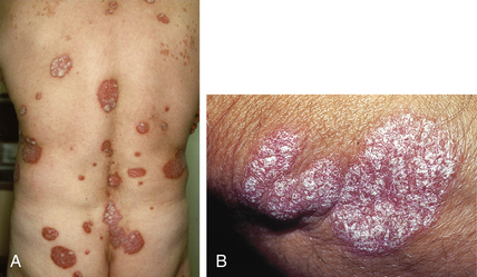
Psoriasis is a common, genetically determined, inflammatory, and hyperproliferative skin disease. Although there are morphologic variations, the most characteristic lesions consist of chronic, well-demarcated, dull-red plaques (Fig. 7-1A) with silvery scale found commonly on extensor surfaces and the scalp (Fig. 7-1B).
4. List the different types of psoriasis.
The different clinical presentations of psoriasis can be separated by morphology or location.
| Morphologic variants | Locational variants |
|---|---|
| Chronic plaque psoriasis | Scalp psoriasis |
| Guttate psoriasis | Palmoplantar psoriasis |
| Pustular psoriasis | Inverse psoriasis |
| Erythrodermic psoriasis | Nail psoriasis |
| Psoriatic arthritis |
5. What is guttate psoriasis?
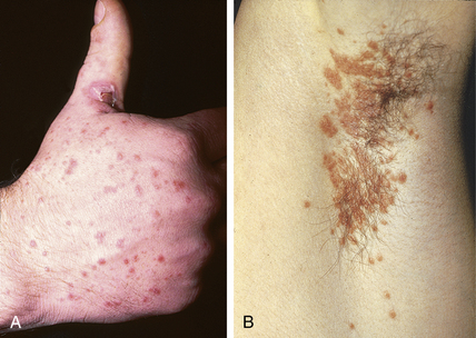
Guttate psoriasis is a variant of psoriasis usually seen in adolescents and young adults. It is characterized by crops of small, droplike, psoriatic papules and plaques (Fig. 7-2A). The word guttate is derived from the Latin gutta, which means “drop.” This type of psoriasis is often found in association with streptococcal pharyngitis, and treatment of the pharyngitis with oral antibiotics may improve or even clear the psoriasis.
6. Does pustular psoriasis refer to psoriasis that is secondarily infected?
No. The pustular forms are uncommon, less stable variants of psoriasis. Instead of erythematous plaques with silvery scale as seen in typical psoriasis, pustular psoriasis is characterized by superficial pustules, often with fine desquamation. Although triggers such as infection can precipitate a flare of pustular psoriasis, the pustules are sterile. Patients often need systemic treatments, such as retinoids, immunosuppressives, or phototherapy, to keep their disease under control.
7. What is inverse psoriasis?
Inverse psoriasis refers to psoriasis that involves intertriginous areas (axillae, groin, umbilicus). This distribution is opposite to the usual extensor distribution of psoriasis vulgaris. Psoriatic lesions with both distributions sometimes can be found in the same patients. Clinically, psoriatic lesions found in these “inverse” distributions often do not have scale, but consist of sharply demarcated red plaques that may become macerated and eroded (Fig. 7-2B). Treatment of inverse psoriasis usually involves low-potency (nonfluorinated) topical corticosteroids.
8. Is there a genetic basis for psoriasis?
Although a specific genetic abnormality has not been identified, psoriasis is generally considered to be a genetically determined disease. There are reports of striking family pedigrees that suggest an autosomal dominant inheritance, but with only partial penetrance. Keep in mind that psoriasis is probably not a single disease, but a family of diseases involving epidermal hyperproliferation. The external environment presumably plays a role in the clinical expression. The strongest evidence for the importance of external factors in the expression of psoriasis is seen in acute guttate psoriasis, which often occurs in association with streptococcal pharyngitis.
9. If one of my relatives has psoriasis, what is the chance that I will get psoriasis?
Questionnaire-based studies reveal that almost 5% of first-degree relatives of psoriasis patients also have psoriasis, compared to 1.2% of relatives of nonpsoriatic spouses of probands. If a sibling has psoriasis, other siblings have a 16% incidence if one parent has psoriasis and a 50% chance if both parents are affected. Twin studies indicate that if one twin has psoriasis, the other twin is similarly affected in 20% of dizygotic pairs and in 73% of monozygotic pairs. The absence of 100% concordance in monozygotic twins (who have identical genetic material) indicates that environmental factors also contribute to the expression of psoriasis.
10. Name the types of psoriatic arthritis.
11. Describe the clinical features of the psoriatic arthritides.
Asymmetrical arthritis, the most common form of psoriatic arthritis, usually involves one or several joints of the fingers or toes. The appearance of this type of arthritis can be similar to subacute gout and include “sausage-like” swelling of a digit due to involvement of the proximal and DIP joints and the flexor sheath (Fig. 7-3). Symmetrical polyarthritis resembles rheumatoid arthritis, but tests for rheumatoid factor are negative, and the condition is clinically less severe than rheumatoid arthritis. Although not common, DIP joint disease of hands and feet is the most classic presentation of arthritis with psoriasis. Destructive arthritis (arthritis mutilans) is a rare, severely deforming arthritis involving predominantly fingers and toes. Gross osteolysis of the small bones of the hands and feet can result in shortening, subluxations, and, in severe cases, telescoping of the digits, resulting in an “opera glass” deformity. Axial arthritis of the spine, which resembles idiopathic ankylosing spondylitis, manifests by itself or with peripheral joint disease.
12. What are the abnormal nail findings seen in psoriasis? Which is most common?
A careful examination of the nails should be part of the skin exam, especially when evaluating a rash that might be psoriasis. Characteristic nail changes are found in 25% to 50% of psoriatics. These changes include nail pitting, discoloration, onycholysis, subungual hyperkeratosis, and nail deformity. Nail pitting, the most common nail finding in psoriasis, consists of small, discrete, punched-out depressions on the nail surface (Fig. 7-4). Circular areas of nail bed discoloration that resemble oil drops are often seen under the nail plate (hyponychium). The nail can become thin and brittle at the distal edge with separation from the nail bed (onycholysis) or thickened with subungual debris. Ridges, grooves, or even frank deformity of the nail plate can also be seen.
Stay updated, free articles. Join our Telegram channel

Full access? Get Clinical Tree


