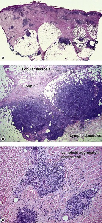
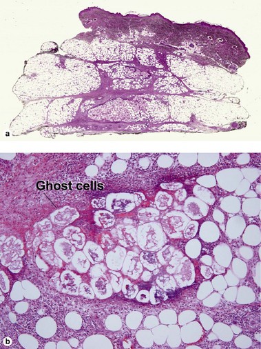
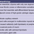
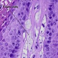
Panniculitis

Chapter 16


Panniculitis


The inflammatory infiltrate in septal panniculitis spills over into the lobule. The inflammation in lobular panniculitis often involves the septum, therefore, septal and lobular panniculitis are best differentiated by the architecture of the lobule. In septal panniculitis, the lobule is intact; lipocytes are similar in size and shape, and the scan appearance resembles a bowl of toasted oat cereal. In lobular panniculitis, the lobule is necrotic; lipophages are common, and in most instances free lipid accumulates in pools that vary in size and shape. The scan appearance resembles the surface of a bowl of chicken soup. Important exceptions to this rule include conditions that result in solidification of fat (sclerema, subcutaneous fat necrosis of the newborn, and steroid-induced fat necrosis). In each of these conditions, the solidified fat cannot coalesce. Pancreatic panniculitis results in calcification of the necrotic membranes, and also prevents coalescence of lipid.


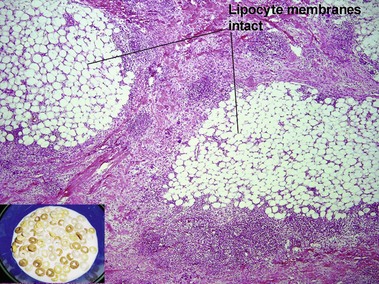
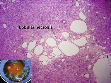
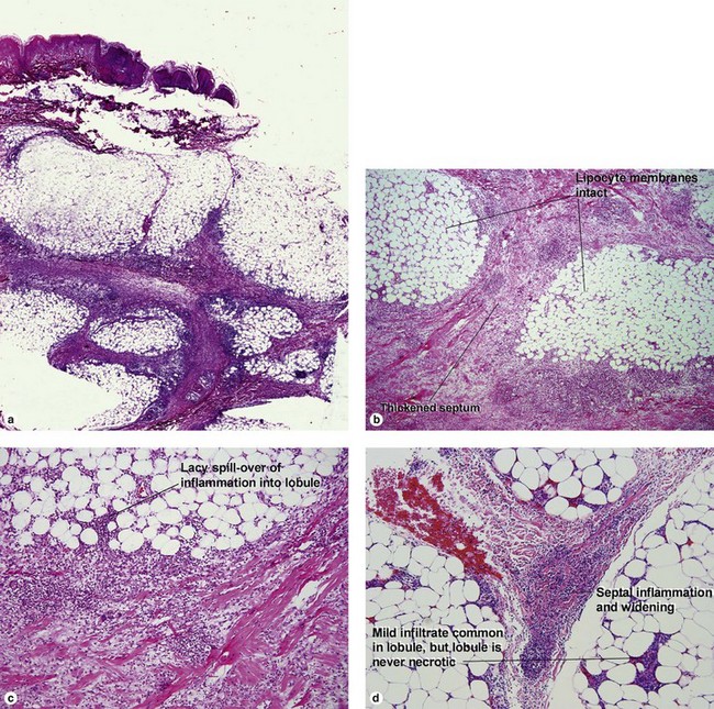
Lobular panniculitis


Lupus panniculitis (lupus profundus)


Although connective tissue disease can produce less specific patterns of granulomatous panniculitis, the features noted above are highly characteristic of lupus panniculitis. Plasma cells are common in the nodular lymphoid foci. Overlying features of lupus erythematosus may be present. The overlying dermis may be sclerotic. Lipomembranous changes may be present (eosinophilic “frost on the windowpane” or “ferning” pattern at the edge of the lipid vacuole).


Apr 26, 2016 | Posted by in Dermatology | Comments Off on Panniculitis

Full access? Get Clinical Tree

Get Clinical Tree app for offline access