(1)
Hôpital Universitaire de Strasbourg, Strasbourg, France
Abstract
Non-purpuric, palpable red lesions are very common and have a great number of causes. They are often the consequence of cutaneous inflammatory diseases. Thus dermatological and extracutaneous associated signs, as well as context, are an important part of differential diagnosis. The “relevant” contexts are numerous and varied: neutropenia and immune deficiency, newly taken drugs, and cold triggering, to name a few. Among possible causes are severe infections (such as septicemias) and cutaneous manifestations in leukemias: the clinical situation of these patients must therefore be carefully analyzed. Indeed, these lesions can easily be trivialized, being very common with very familiar causes, such as insect bites or urticaria.
Non-purpuric, palpable red lesions are very common and have a great number of causes. They are often the consequence of cutaneous inflammatory diseases. Thus dermatological and extracutaneous associated signs, as well as context, are an important part of differential diagnosis. The “relevant” contexts are numerous and varied: neutropenia and immune deficiency, newly taken drugs, and cold triggering, to name a few. Among possible causes are severe infections (such as septicemias) and cutaneous manifestations in leukemias: the clinical situation of these patients must therefore be carefully analyzed. Indeed, these lesions can easily be trivialized, being very common with very familiar causes, such as insect bites or urticaria.
Certain papular inflammatory dermatoses can be clinically identified, for example, typical lichen papules. In such a case, they have a characteristic purple coloration and are flat, polygonal, and covered with fine white striations, called “Wickham’s striae.” They are pruritic. In case of lesions of secondary syphilis, a perilesional collarette surrounding the papule is very suggestive of this diagnosis, especially when lesions are located on extremities.
Neutrophilic dermatoses (i.e., Sweet syndrome), vasculitides, and erythema multiforme are possible causes of multiple, eruptive, edematous plaques.
Inflammatory lesions located in the hypodermis (deep-seated nodules, or “nouures” in French terminology) reflect an affection either of the adipose lobules (lobular panniculitis), of the inter-adipose septa (septal panniculitis), or of hypodermal vessels (either veins or arteries).
Papules often have an altered surface and produce intricate lesions which are discussed later on, i.e., papulopustules, scaly papules, and necrotic papules.
Box 35.1
Main Causes of Erythematous Papules
Acne |
Atypical fibroxanthoma |
Botryomycoma |
Burn caused by marine animals and plants |
Chilblain |
Cutaneous Kikuchi disease |
Cutaneous manifestations in septicemias |
Eosinophilic folliculitis (usually pustular) |
EPPER (eosinophilic, polymorphic, and pruritic eruption associated with radiotherapy) |
Eruptive xanthoma (pink) |
Erythema elevatum diutinum (on the back of the hands) |
Fibroblastic rheumatism |
Flea bites |
Folliculitis caused by infection with Malassezia (usually pustular) |
Foreign body granuloma |
Gianotti-Crosti syndrome |
Gluteal granuloma |
Gottron’s papules in dermatomyositis (back of the hands) |
Granuloma annulare (which generally merges into annular lesions) |
Grover’s disease (often papulovesicular) |
Hordeolum (or sty) and chalazion |
Insect bite and sting |
Kaposi’s disease |
Lymphomatoid papulosis |
Multicentric reticulohistiocytosis (back of the hands) |
Perforating elastoma (that merge into arciform lesions) |
Pityriasis lichenoides |
Prurigo |
Rickettsioses |
Rosacea |
Ruby spots (or cherry angioma) |
Sarcoidosis (often annular) |
Scabies |
Secondary syphilis |
Urticaria |

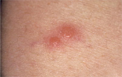
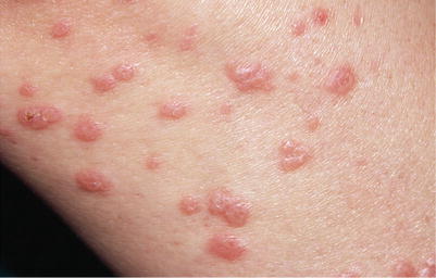
Fig. 35.1
Succulent (i.e., “impregnated with fluid”) erythematous papules on an erythematous macule. Prurigo


