(1)
Hôpital Universitaire de Strasbourg, Strasbourg, France
Abstract
There are several types of palpable lesions that can be identified according to their content (fluid or solid), the possible presence of an alteration of the surface of the skin, and their size and location within the skin (superficial or deep).
There are several types of palpable lesions that can be identified according to their content (fluid or solid), the possible presence of an alteration of the surface of the skin, and their size and location within the skin (superficial or deep).
The mechanisms underlying these lesions can be determined by the sole histopathological examination: edema, infiltration by inflammatory or tumoral cells, and substance overload (amyloidosis, mucinosis, etc.).
4.1 Papule
The papule is usually defined as a small palpable lesion, not exceeding 10 mm, with non-fluid content (Fig. 4.1). In the USA and the UK, the maximum width of a papule is restricted to 5 mm in many textbooks. Lesions are generally elevated, above the level of the adjacent skin. Seen from above, a papule may be round (Fig. 4.2), oval, umbilicated (with a small central depression) (Fig. 4.3), or polygonal. Seen in profile, it can be flat, domed, sessile, pedunculated, or acuminate (conical, pointed) (Fig. 4.4). The surface may be either smooth (Fig. 4.5), eroded, ulcerated, or necrotic (Fig. 4.6), covered with scales (Fig. 4.6), crusts, or scaly crusts (Fig. 4.6); hair follicles may be prominent and confer a “peau d’orange” (orange peel) appearance (Fig. 4.7). Finally the distribution pattern may or may not be follicular (Fig. 4.4).
The consistency of a papule varies depending on its nature; some lesions are very hard, others soft and depressible (Fig. 4.8). Papules that result from epidermal proliferation or deposits (e.g., plane wart, seborrheic keratosis) usually have clear-cut and rectilinear boundaries and are often rough (Fig. 4.9), whereas dermal papules (e.g., granuloma annulare) are smoother and borders are less defined (Fig. 4.10). Papules must be distinguished from other palpable lesions which can be larger (plaques, nodules, tumors), fluid-filled (vesicles, bullae), or mainly due to alteration of the lesion’s surface (cutaneous horn, keratosis), which are also called keratotic papules.
The term tuber should not be used. It is a palpable intradermal lesion, either flat or barely elevated. Therefore, it is merely an intradermal papule (Fig. 4.11). These lesions often become chronic or tend to leave a scar on regression (e.g., lupus vulgaris). They are circumscribed and movable, relatively to the hypodermis.
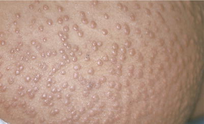
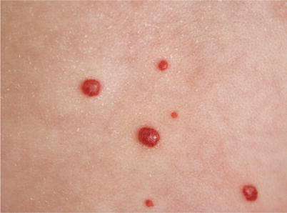
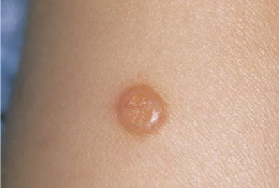
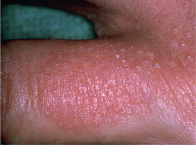
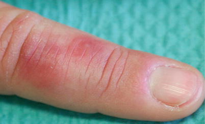
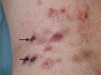
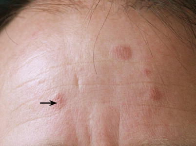
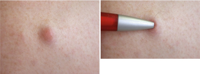
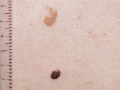
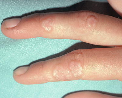
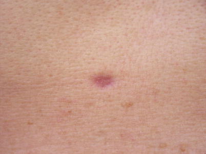
Fig. 4.1
Papules. Eruptive xanthomas. They consist of papules, i.e., palpable red or orange-yellow lesions of less than 1 cm (0.5 cm in the USA), showing no deterioration of the skin surface. They are usually numerous and of sudden onset. They are the cutaneous expression of an often major hypertriglyceridemia that can be life threatening due to the risk of pancreatitis
Fig. 4.2
Multiple hemispherical, dome-shaped, angiomatous papules. Diffuse neonatal hemangiomatosis. This is a potentially serious disorder because of the likelihood of visceral hemangiomas, particularly in the liver and spleen, high-output heart failure, and hypothyroidism caused by impaired secretion of a thyronine-degrading enzyme
Fig. 4.3
Umbilicated papule. Molluscum contagiosum. Hemispherical pink papule, with a central depression called umbilication, highly characteristic of a virus-induced lesion. Telangiectasia may also be identified laterally. Molluscum contagiosum is a lesion related to an infection with a poxvirus. It is frequent in children. When occurring in great numbers in adults, it is essential to search for an immune deficiency
Fig. 4.4
Acuminate keratotic papules (pilar papule), on an erythematous plaque. Pityriasis rubra pilaris. These papules are cone-shaped, with a superficial apex and an epidermal base, typical of a pilar papule
Fig. 4.5
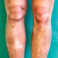
Red, smooth, edematous papules, without alteration of the skin surface. Chilblain (pernio). These papules are poorly defined and erythematous. Chilblain is characterized by its location on extremities and its onset in cold and humid conditions. It is mostly idiopathic and can also reveal disorders such as myelomonocytic leukemia, lupus erythematosus, antiphospholipid antibody syndrome, cryopathy, or Aicardi-Goutieres syndrome in infants
Fig. 4.6
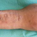
Papules and necrotic plaques. Lymphomatoid papulosis. These papules and red plaques become scaly, crusty, and necrotic (arrows). They are covered with hemorrhagic crust. This is a typical progression of lymphomatoid papulosis, an auto-abortive lymphoma conferring an increased risk of Hodgkin’s disease, anaplastic lymphoma, and mycosis fungoides
Fig. 4.7
Erythematous papules. Lever’s granuloma. Note the protruding hair follicles conferring a “peau d’orange” (orange peel) appearance (arrow) characteristic of this disorder, mostly located on the face
Fig. 4.8
Depressible exophytic papule. Neurofibroma. A papule is rarely depressible. It is particularly found in neurofibroma, anetoderma, certain nevi, piezogenic papules, and pendulum
Fig. 4.9
Dark brown papule and light brown plaque. Seborrheic keratoses. Note the highly rectilinear boundaries; these two lesions seem “laid” on the skin. This is characteristic of epidermal papules. Compare the borders with those of the lesion on Fig. 4.10. Seborrheic keratosis is the result of proliferation of keratinocytes, the cells of the epidermis. It is a benign and very common lesion, especially in patients over 60 years old
Fig. 4.10
Papules and plaques in annular configuration. Granuloma annulare. Note the borders of this lesion: they are less rectilinear and less clear-cut than those of Fig. 4.9. The skin appears lifted by an underlying lesion. This semiological aspect characterizes dermal lesions; i.e., at the microscopic level, granuloma annulare is a granulomatous inflammation of the dermis (“palisading granuloma”)
Fig. 4.11


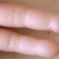
Intradermal papule. Fibroma. The lesion has a very firm consistency on palpation. This papule is depressed below the skin surface. Pinching between thumb and forefinger forms small cavities which partially cover the lesion (dimple sign). This sign is characteristic of fibroma
Related posts:

Stay updated, free articles. Join our Telegram channel
Full access? Get Clinical Tree
