Rhinoplasty is one of the oldest and most commonly performed facial plastic surgery procedures. It is an operation performed by surgeons of all skill levels with different training backgrounds. As such, it is easier to group all such surgeons who perform this operation into the category of “rhinoplastic surgeons.”
No matter what the background or skill level of the rhinoplastic surgeon, most will undoubtedly agree that rhinoplasty is a relatively difficult operation to grasp early on in one’s career during residency and that it takes years to gain mastery of the nuances of the procedure. One gains a true sense of the intricacies of the operation after studying the work of the various surgeons who have dedicated their entire careers to this operation. To hear these giants in the field admit humility and see how they have learned from and fine tuned their results after 30 years or more of experience is an opportunity not to be missed or taken lightly.
Even after years of training and years of fine tuning technique, at times each surgeon will encounter a difficult nose or end up with results that are less than satisfactory to the patient or surgeon. Just as a less-than-harmonious unoperated nose attracts undue attention and may have an adverse psychosocial effect on a patient, an operated nose, even when greatly improved from the presurgical state, is placed under scrutiny by the patient, the likes of which were not present preoperatively. Every subtle irregularity is now highlighted and noticed.
When encountering subtle irregularities or imperfections in the immediate postoperative period, whether our own patient or a patient operated on by a colleague, the initial prudent technique is that of patient education and patience. Seldom can an operated nose be improved by haste in the healing period.
For a variety of reasons, a surgeon may need to operate on a nose that has been operated on previously, either by the same surgeon or another. Often, this may be the third or fourth operation, making the term “revision rhinoplasty” possibly more descriptive than the commonly used term, “secondary rhinoplasty.” A variety of reasons contribute to the need for revision rhinoplasty. These include, but are not limited to, poor surgical planning, improper technique, underresection or more commonly overzealous reduction rhinoplasty, very thick or very thin nasal–soft tissue envelope, insufficient nasal framework, unpredictable healing, inadequate surgeon and patient preoperative communication, unrealistic patient expectations, or traumatic injury to the previously operated nose.
Revision rhinoplasty introduces a new series of challenges for the facial plastic surgeon. Variable degrees of scarring, loss of nasal support mechanisms from aggressive reduction rhinoplasty, and lack of adequate septal cartilage for rebuilding, are only some of the obstacles a surgeon may face venturing back into a previously operated nose. The use of auricular cartilage or other suitable building blocks, such as rib cartilage, irradiated cartilage, Gore-Tex Subcutaneous Augmentation Material (GORE S.A.M., W. L. Gore & Associates, Inc., Flagstaff, AZ), AlloDerm (LifeCell, Branchburg, NJ) or other acellular tissue, and other alternatives to autogenous septal cartilage are also more common than in primary rhinoplasty. However, even in secondary rhinoplasty, allografts should be used as an alternative rather than a substitute for the more preferred autografts.1 A graft material not commonly used but worth consideration is autologous dermal graft especially for patients concerned about the potential of prions and other small infectious particles possibly associated with cadaveric tissue.2
Preoperative planning, including in-office patient exam and counseling, is a crucial investment of time. We cannot stress enough the importance of “imaging.” This is an opportunity for the surgeon and patient each to communicate visually their respective goals for the operation. This technology also allows the surgeon to show the possible limitations of the operation with respect to each patient’s anatomy through the use of morphing software. The office consult provides a forum for the discussion of possible implant choices. The recovery room is not the ideal place to inform a patient that he or she now has a foreign or cadaveric implant if this possibility had not been previously addressed with the patient. Yet each patient must be aware that it is usually after entering the nose that the surgeon can properly evaluate what was previously done and what further needs to be done to correct the problem. The columellar incision must also be mentioned to the patient. More often than not, major revisions, especially of the lobule, will necessitate an external approach, whereas other problems may be approached through an endonasal technique for pocket grafting, alar retraction correction, or dorsal refinement.
 Special Problems
The problems requiring revision rhinoplasty can be categorized in relation to the anatomic site as well as the types of aesthetic and functional defects commonly seen. Common areas to address include the pyramid, lobule, and airway. Most of these issues can be attributed to errors of “omission” or errors of “commission.” We define errors of “omission” as those maneuvers that needed to be done and were not in the previous surgery. On the contrary, errors of “commission” are those maneuvers that were not necessary in the previous surgery or were done too aggressively, leaving the nose usually destabilized with an overoperated appearance. In this chapter, we will present the most common reasons for revision rhinoplasty in our practice and offer some time-tested solutions to add to your surgical armamentarium.
Errors of Omission
Errors of omission most commonly include inadequate tip refinement, dorsal hump reduction, or pyramid narrowing. A nose that is still overprojected or underrotated is yet another example of this error. These problems are easy to address and require completion of the maneuvers that were either done too conservatively in the previous operation or not done at all (Fig. 20–1).
Here and elsewhere throughout this chapter, you will appreciate that the first step in correction of any nasal deformity, whether primary or revision, is the appropriate diagnosis of the internal structural variations leading to the external aesthetic or functional abnormality. As in any area of medicine and surgery, diagnosis is the initial, crucial step. The good rhinoplasty surgeon studies each nose, diagnoses the problem, and offers a tailored solution. Far too frequently, surgeons learn a “standard” rhinoplasty operation and apply the same series of maneuvers to each nose, regardless of the problem at hand and the subtle individual variations in anatomy. Without the appropriate diagnosis, the proper surgery cannot be performed.
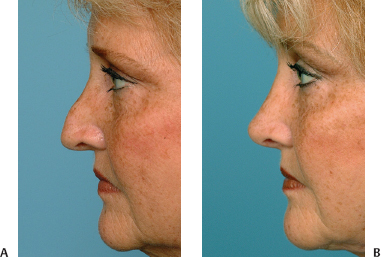
Figure 20–1 “Undersurgery” by another surgeon resulted in a classic pollybeak deformity, which was corrected mainly through completion of dorsal cartilage resection. (A) Preoperative and (B) postoperative photos.
The Overprojected Tip
There are multiple causes of an overprojected tip and hence multiple techniques for addressing this problem. These techniques include excess length of the caudal septum, long lower lateral cartilages, a “hanging” or underrotated tip giving the appearance of overprojection, and previously excessive augmentative use of tip grafts. It is crucial to realize the aesthetic relationship between tip projection and rotation and how each surgical maneuver may affect one or both. Our first choice for deprojection is a complete transfixion incision, disrupting nasal tip support mechanisms. The second maneuver would be appropriate resection of the caudal septum. If further deprojection is needed, the Lipsett technique is used; we use 6.0 PDS sutures for this purpose. This technique involves transection of the medial crus of the lower lateral cartilage somewhere between its upper and middle third, followed by overlapping and suturing to shorten the medial crus of the lower lateral cartilage. In addition to deprojection, this maneuver creates derotation. Although usually done bilaterally, the Lipsett technique can be done unilaterally to correct tip asymmetries. The original description by Lipsett did not include suture stabilization, but we believe given the contracture caused by healing, suturing allows for more predictable results.3
The Underrotated Tip
Related posts:
Stay updated, free articles. Join our Telegram channel

Full access? Get Clinical Tree


