Chapter 22. Open Approach to the Overprojecting Tip
C. Spencer Cochran, MD; Jack P. Gunter, MD
INDICATIONS
An incremental approach to tip deprojection in open rhinoplasty is indicated for patients who have an overprojecting nasal tip. Factors that contribute to tip support, and thus tip projection, include: (a) the length and strength of the lateral crura; (b) the length and strength of the medial crura; (c) the fibroelastic attachments connecting the feet of the medial crura to the caudal septum; (d) the ligamentous attachments between the lateral crura and the upper lateral cartilages; (e) the suspensory ligament over the septal angle connecting the cephalic margins of the domes; (f) the septal angle; and (g) the nasal skin.
PREOPERATIVE PREPARATION
A thorough preoperative nasal examination and analysis of preoperative photographs should be performed. Using computer morphing software or tracing paper enables the surgeon to estimate the planned tip projection. If preoperative analysis indicates that one goal of rhinoplasty is to decrease tip projection, the surgeon needs to be able to control the decrease incrementally.
ANESTHESIA
General anesthesia with endotracheal intubation is preferred for this procedure. This allows the patient to be completely still for the duration of the procedure and allows for a protected airway.
POSITION AND MARKINGS
The patient is placed in the supine position. After adequate anesthesia is administered, the patient’s nasal passages are packed with one-inch gauze strips saturated with a topical decongestant such as oxymetazoline or phenylephrine. The soft-tissue envelope is injected with approximately 5 mL of 1% lidocaine with 1:100,000 epinephrine, and the patient is prepped and draped in a sterile fashion.
The planned transcolumellar incision is marked as an inverted V or chevron shape across the narrowest portion of the columella and extends around the columellar roll to terminate on the midportion of the medial crura.
DETAILS OF PROCEDURE
Increased tip projection may be a result of any one of the aforementioned factors; usually it is the result of a combination of the factors, and therefore requires a stepwise or incremental approach. After each factor is addressed, the effect on tip projection should be assessed by redraping the soft-tissue envelope.
The transcolumellar incision is created with a no. 15 blade scalpel and connected with infracartilaginous incisions that follow the caudal border of the lateral crura. The nasal soft-tissue envelope is elevated from the tip and nasal dorsum to expose the osseocartilaginous framework. The soft-tissue envelope should be widely undermined from the lateral crura toward the piriform aperture because destabilization of the nasal tip occurs when the soft-tissue envelope is elevated in the open approach, allowing for a modest amount of deprojection.
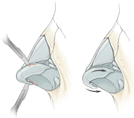
Sever the ligamentous attachments between the lateral crura and the upper lateral cartilages (Fig. 22-1) and divide the suspensory ligament traversing the septal angle that connects the cephalic margins of the domes (removal of the cephalic margin of the lateral crura has the same effect). After the suspensory ligament has been divided, patients with a prominent septal angle may require judicious trimming of the lower portion of the cartilaginous dorsal septum. This can help prevent a “polly-beak” deformity resulting from excess dorsal septum in the supratip area.

Figure 22-1 Decreasing tip projection by release of the ligamentous attachments between the lateral crura and upper lateral cartilages and release of the fibroelastic attachments between the medial crura and caudal septum.
Stay updated, free articles. Join our Telegram channel

Full access? Get Clinical Tree


