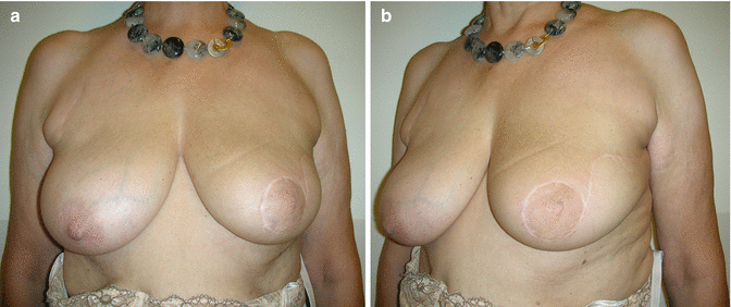
Fig. 33.1
(a, b) Preoperative view. The 54-year-old patient had a 22 mm invasive breast cancer and DCIS in the upper outer quadrant of the left breast. Preoperative drawings for B plasty
33.2 Surgery
The tumor was palpable in the upper outer quadrant of the left breast. Due to concomitant intraductal carcinoma in situ, a wide resection was planned using a B plasty (Fig. 33.1a, b). The area around the areola was de-epithelialized. Quadrantectomy was done with a wedge of tissue resected underneath the nipple-areola complex. The tissue around the areola was incised to facilitate rotation of the flaps for closure. The breast flaps were adequately mobilized from the major pectoralis muscle fascia and sutured together. Sentinel lymph node biopsy was done through the same incision and found two negative nodes.
33.3 Clinical and Cosmetic Outcome
The permanent histological examination found a 22 mm invasive cancer and a 30 mm intraductal carcinoma of intermediate grade which were both removed with wide free margins. The postoperative course was uneventful. Postoperative radiation and endocrine treatment were suggested. The postoperative cosmetic result was rated as excellent by both the patient and the surgeon (Fig. 33.2a, b).


