EVALUATION OF THE EYELIDS
John L. Wobig and Roger A. Dailey
HISTORY
A thorough history taken from the patient or family is essential. A routine eye examination is fundamental and should include vision, external examination, gross confrontation fields, a tear film analysis, extraocular movements, slit lamp exam, and fundus exam if indicated. A detailed medication list and allergy list determines your selection of surgery and anesthesia. Past history is important to determine previous surgical procedures, radiation, and drug therapy. Family history of preexisting disease may give diagnostic clues to patients with eyelid disease.
Determining the patient’s main concern is paramount to a successful surgical outcome. A technically spectacular result won’t matter if it does not correct what the patient is concerned about. Use of a mirror in the office allows patients to point out the features they want corrected.
Table 3-1 lists important historical information that should be elicited from all patients. Specific problems to be aware of are angina, orthopnea, thyroid abnormalities, allergies, previous anesthetic complications, and previous eyelid surgery. Use and type of contact lenses should be noted. Social history often provides insight into the patient’s reasons for seeking surgery and may be used to justify surgery for functional reasons (e.g., a visual field deficit that interferes with job performance). The patient should be questioned carefully about bleeding tendencies, and any platelet inhibitors or anticoagulants should be discontinued prior to surgery. Notification of the patient’s primary care provider is not only appropriate as a courtesy, it may also be required for prior authorization in a managed care environment.
| Age | |
| Medical history | |
| Past surgeries | |
| Ocular history | |
| Family history | |
| Social factors | |
| Review of systems | |
| Medications | |
| Allergies | |
| Bleeding tendencies | |
| Primary care physician |
EXAMINATION
Ideally, the examination begins the moment the physician enters the room. Grooming, head posture, face turn, habitual brow elevation, skin texture, facial contours and symmetry, hairline position, and ocular motility disturbances are all helpful to note. Many are best observed prior to the patient’s realization that the exam has begun.
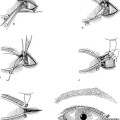
Prior to any direct measurements, observe facial symmetry, protractor and contractor function, and location of lesions. In ptosis patients look for unilateral or bilateral ptosis, presence or absence of a crease, and levator function. In patients with eyelid lesions observe the location and presence of singular or multiple lesions.
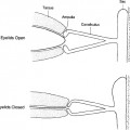
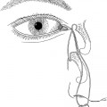
Table 3-2 lists the major information to be documented in examining the eyes and eyelids. It is very important to document preoperative visual acuity and health of the eye. If postsurgical complications develop, documentation of antecedent ocular conditions helps determine which problems may be associated with the surgery and can help avoid serious medicolegal problems. Corneal sensation testing, slit lamp examination, fluorescein staining, and Schirmer’s basic tear secretion give the examiner an idea of the health and stability of the cornea. Because topical steroids are often used in combination with an antibiotic postoperatively, documentation of preoperative intraocular pressure (IOP) is wise and functions as a useful glaucoma screen.
TABLE 3-2 PHYSICAL EXAMINATION
| Ocular examination | |
| Visual acuity | |
| Pupils | |
| Extraocular movements (Bell’s) | |
| Gross confrontational fields | |
| Alternate cover testing | |
| Corneal sensation | |
| Schirmer’s basic secretion | |
| Slit lamp | |
| Fluorescein dye | |
| Applanation tonometry | |
| Eyelid examination | |
| Brow position | |
| Skin | |
| Orbital fat pads |