(1)
Department of Neurosurgery Faculty of Medicine, Saga University, Saga, Japan
Keywords
Bypass surgeryOccipital artery–posterior inferior cerebellar artery anastomosisOccipital arteryPosterior inferior cerebellar arteryVertebrobasilar insufficiencyVertebral artery–posterior inferior cerebellar artery aneurysm21.1 Introduction
An occipital artery–posterior inferior cerebellar artery (OA–PICA) anastomosis is performed when PICA is sacrificed during the treatment of poor posterior circulation. Ausman JI, et al. [1] performed the first intracranial posterior circulation revascularization procedure for vertebrobasilar insufficiency in the form of an OA–PICA anastomosis in 1976. Sundt TM Jr. and Piepgras DG [12] and Khodadad G [5] reported additional experiences of this procedure. Since then, OA–PICA anastomosis has been important for cerebral revascularization in the posterior circulation [2, 6, 10, 11]. Although the benefits of intracranial OA–PICA bypass surgery for vertebrobasilar insufficiency caused by atherosclerotic disease of the extracranial or intracranial vertebral artery (VA) remain unclear, cerebral revascularization of the posterior circulation is well established for the treatment of complex and giant intracranial aneurysms and tumors, especially when these lesions involve the major vessels of the posterior circulation.
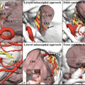
Three factors contribute to a successful OA–PICA bypass surgery. First, an adequate dissection of OA is necessary. Second, a wide operative field at the anastomotic site below the cerebellar tonsil is required for a safe procedure; therefore, skull base surgery techniques, such as the far lateral [3] and transcondylar fossa [8] approaches, are required. In some cases, the cerebellomedullary fissure (CMF) is opened [9]. Third, accurate dural closure without disturbing the bypass flow is mandatory.
21.2 Microsurgical Anatomy
21.2.1 The Occipital Artery
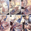
OA arises from the posterior portion of the external carotid artery, opposite the external maxillary artery and near the lower margin of the posterior belly of the digastric muscle, and ends in the posterior portion of the scalp. At its origin, OA is covered by the posterior belly of the digastric muscle and the stylohyoid muscles. Cranial nerve (CN) XII winds around this artery from behind forward. OA crosses the internal carotid artery, internal jugular vein, and CNs X and XI. It then ascends between the transverse process of the atlas and the mastoid process of the temporal bone, and passes horizontally backward, grooving the surface of the mastoid process, while covered by the sternocleidomastoid, splenius capitis, longissimus capitis, and digastric muscles and resting on the rectus capitis lateralis, obliquus capitis superior, and semispinalis capitis muscles. The mean diameter of OA is 2.05 mm at its exit from the digastric groove [4]. There, OA changes course and runs vertically upward, piercing the fascia connecting the cranial attachment of the trapezius and sternocleidomastoid muscles, and ascends in a tortuous course in the superficial fascia of the scalp, where it divides into numerous branches that reach as high as the vertex of the skull and anastomose with the posterior auricular artery and superficial temporal artery (STA). The mean diameter of OA is 2.01 mm at the level of the superior nuchal line, and the mean length of this segment is 81.9 mm [4]. Its terminal portion is accompanied by the greater occipital nerve (Figs. 21.1 and 21.2).
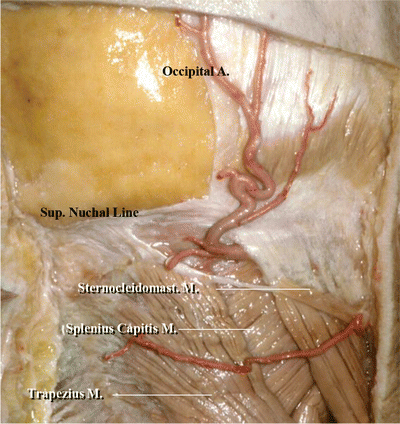
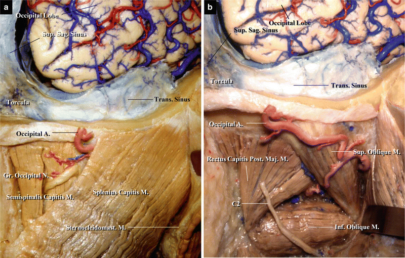
Fig. 21.1
The mean diameter of the occipital artery (OA) was 2.05 mm at its exit from the digastric groove. The mean diameter of OA was 2.01 mm at the level of the superior nuchal line. The mean length of this segment was 8.19 cm. Its terminal portion was accompanied by the greater occipital nerve (from Kawashima M [4] with permission)
Fig. 21.2
The occipital artery changes course and runs vertically upward at its exit from the digastric groove, pierces the fascia connecting the cranial attachment of the trapezius and sternocleidomastoid muscles, and ascends in a tortuous course in the superficial fascia of the scalp, where it divides into numerous branches that reach as high as the vertex of the skull (from Kawashima M [4] with permission)
21.2.2 The Posterior Inferior Cerebellar Artery
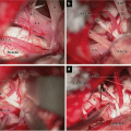
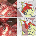
PICA is divided into five segments: anterior medullary, lateral medullary, tonsillomedullary, telovelotonsillar, and cortical [7]. The tonsillomedullary segment, which forms a caudally convex loop called the caudal loop, is usually selected for a recipient artery. When the tonsillomedullary segment is not suitable, the telovelotonsillar segment is sometimes utilized after opening CMF (Fig. 21.3). (Regarding the anatomy of PICA, refer to Chap. 3: “Three Cerebellar Arteries: Superior Cerebellar Artery, Anterior Inferior Cerebellar Artery, and Posterior Inferior Cerebellar Artery.”)
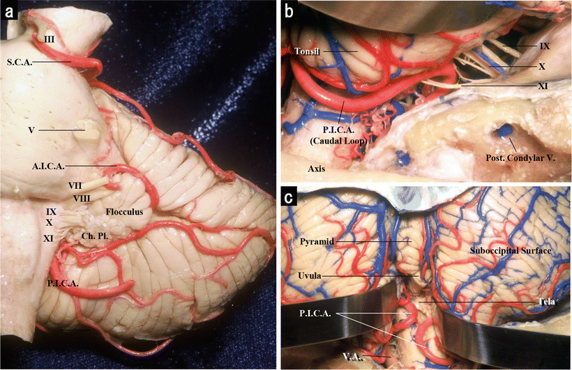
Fig. 21.3
(a) Anterior view of the left brainstem and cerebellum. The posterior inferior cerebellar artery (PICA) arises from the vertebral artery near the inferior olive and passes posteriorly around the medulla oblongata. (b) The right cerebellomedullary cistern below the right cerebellar tonsil. After PICA passes the lateral aspect of the medulla, it courses around the cerebellar tonsil, enters the cerebellomedullary fissure, and passes posterior to the lower half of the fourth ventricle roof. The portion passing near the lower part of the tonsil, termed the caudal loop, comprises a caudally convex loop that coincides with the caudal pole of the tonsil (from Kawashima M [4] with permission). (c) The telovelotonsillar segment. The telovelotonsillar segment begins beyond halfway across the medial surface of the tonsil toward the roof of the fourth ventricle and ends at the point at which PICA exits the fissures between the vermis and tonsil on one side and the hemisphere on the other side to reach the suboccipital (posterior) cerebellar surface. This segment commonly forms a loop with a convex rostral curve, called the cranial loop (from Kawashima M [4] with permission)
21.3 Surgical Procedure for OA–PICA Anastomosis
21.3.1 Patient Position
The patient is usually placed in a prone position, although a park bench position, placing the lesion side upward, is an alternative.
21.3.2 Skin Incision
The course of OA is outlined on the scalp using a portable Doppler device. A unilateral, horseshoe-shaped skin incision is performed. OA is cut, but left intact at the level of the rostral margin of the skin incision.
21.3.3 Dissection of OA
Related posts:
 The Veins of the Posterior Cranial Fossa: Nomenclature
 The Cerebellopontine Angle: Basic Structures and the “Rules of Three”
 Microvascular Decompression Surgery for Hemifacial Spasm: The Lateral Suboccipital Infrafloccular Approach
 Microvascular Decompression for Glossopharyngeal Neuralgia: Surgical Approaches Depending on the Offending Artery
 Microsurgical Anatomy of and Surgical Approaches to the Jugular Foramen
 Microsurgical Anatomy of the Cerebellomedullary Fissure and Variations of the Transcerebellomedullary Fissure Approach
Stay updated, free articles. Join our Telegram channel

Full access? Get Clinical Tree


