Fig. 44.1
(a–c) The 47-year-old BRCA 1-positive patient was diagnosed with a 12 mm invasive cancer in the right breast. Nipple-sparing, skin-reducing bilateral mastectomy was planned with immediate reconstruction with implants and an ADM. The breast was large and ptotic
44.2 Surgery
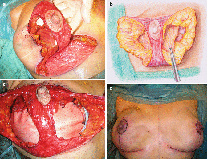
Nipple-sparing skin-reducing bilateral mastectomy was performed with a resection weight of 710 g for the right breast and 680 g for the left breast. The areola was circumcised and a superior pedicle and an inferior pedicle were de-epithelialized. The mastectomy was done through a vertical incision lateral of the inferior pedicle with a flap thickness of a few millimeters (Fig. 44.2a).


Fig. 44.2
(a–d) The mastectomy was performed leaving thin de-epithelialized superior and inferior flaps for blood supply of the nipple (a). The de-epithelialized inferior pedicle further supports the implant and should provide adequate blood supply to the nipple, but may be removed in case too much breast tissue is left behind. The insertions of the pectoralis major muscle were dissected off its origin in the inframammary fold (b). The implants were inserted in the subpectoral pocket and covered with an ADM (c) in the inferior pole and the wound closed in an inverted T pattern (d)
The major pectoral muscle was dissected off its origins in the inframammary fold and caudomedially (Fig. 44.2b), and a 410 cc anatomical implant was inserted in the subpectoral pocket and covered with an acellular dermal matrix (ADM) (Fig. 44.2c


Stay updated, free articles. Join our Telegram channel

Full access? Get Clinical Tree


