Fig. 42.1
(a, b) Preoperative view. The 46-year-old patient had a multicentric lobular cancer of the right breast. The incision for the nipple-sparing mastectomy was horizontal in the lateral quadrant. The breast was of medium size and moderate ptosis
42.2 Surgery
A nipple-sparing mastectomy was done using a horizontal incision in the lateral breast. Intraoperative frozen section biopsy found no tumor in the retroareolar biopsy and the nipple was preserved. The resection weight of the mastectomy specimen was 570 g.
The insertions of the pectoralis major muscle were dissected off the inframammary fold and medially to the height of the nipple in a standing position. A 420 cc anatomical implant was inserted under the pectoralis major muscle, and the distance between the inframammary fold and the caudal insertions of the pectoralis major muscle was covered with an acellular dermal matrix (ADM).
Sentinel node biopsy was done through an axillary incision and revealed two negative sentinel nodes. Two drains were placed in the submuscular and mastectomy pocket. Perioperative antibiotics were given perioperatively and then for 3 days after surgery. One drain was removed 4 days after surgery and the patient was discarded with the second drain in place.
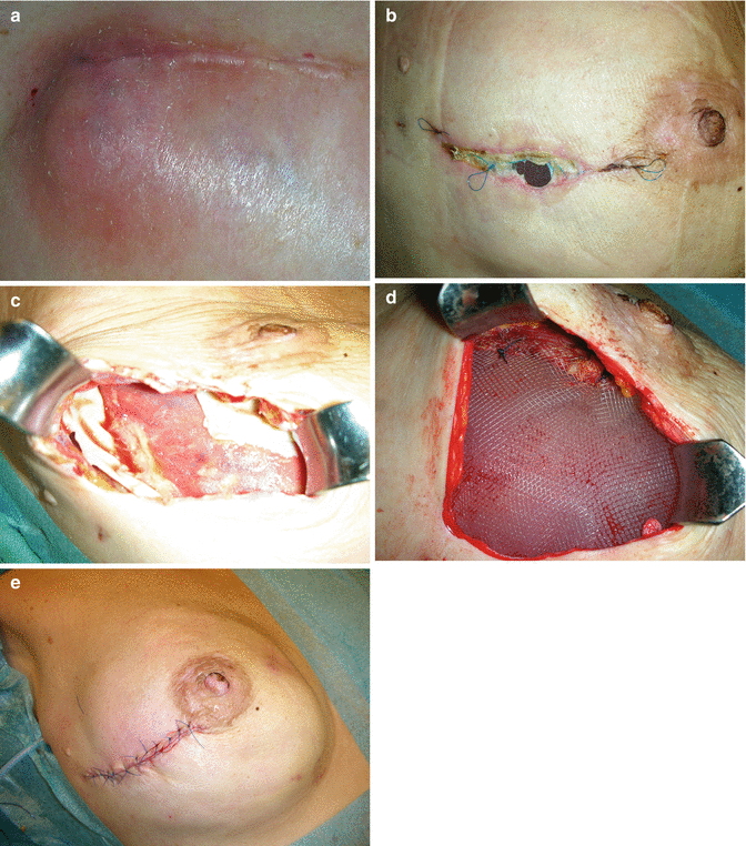
Early postoperative course was uneventful. On the tenth day following surgery, a small reddening was seen in the area of the incision and increased over the following days despite antibiotic coverage (Fig. 42.2a). White blood cell count was normal; the patient showed no fever or any other signs of local or generalized infection. No local necrosis of the mastectomy flaps was seen and there was no seroma as well. The reddening slowly decreased over the next days, and a couple of days later, local breakdown of the scar and exposure of the implant were seen (Fig. 42.2b).


Fig. 42.2
(a–e) The local reddening and swelling of the skin (a) was followed by breakdown of the wound and exposure of the implant (b). There was no coverage of the implant with the ADM. Intraoperatively the ADM was dissolved over the implant and only small parts were attached at the pectoralis major muscle and the serratus fascia (c). The implant was covered with an extralight TiLOOP Bra mesh fixed to the muscle (d) and the wound was closed (e)
The patient was scheduled for reoperation surgery. The ADM was found completely dissolved over the implant and only partially attached to the pectoralis major muscle and the fascia of the serratus anterior muscle (Fig. 42.2c


Stay updated, free articles. Join our Telegram channel

Full access? Get Clinical Tree


