(1)
Department of Neurosurgery Faculty of Medicine, Saga University, Saga, Japan
Keywords
Neural structureBrainstemCerebellumCerebellar pedunclesFourth ventricle2.1 Introduction
The neural structures of the posterior cranial fossa include the brainstem and cerebellum, which are connected by the cerebellar peduncles. Together, they form the fourth ventricle. Three deep cerebellar–brainstem fissures exist beneath the three cerebellar surfaces [14, 15]. These structures can be visualized by magnetic resonance imaging [7, 8, 11]. To understand the neural structures of the posterior cranial fossa, neurosurgeons must first study the structures that must be navigated to reach the fourth ventricle. In this chapter, an explanation of the basic anatomy of the fourth ventricle will be followed by a description of the cerebellar surfaces. The relationship between the cerebellar surfaces and fourth ventricle will then be elucidated to demonstrate the surgical approaches to the fourth ventricle. Because the floor of the fourth ventricle is a challenging area for neurosurgeons, its surgical anatomy will be explained in detail. The anatomy of the three cerebellar peduncles in relation to surgical approaches to intrinsic lesions of the brainstem will also be introduced.
2.2 Basic Anatomy of the Fourth Ventricle
The floor of the fourth ventricle is formed by the pons and medulla oblongata. The rostral two-thirds of the floor is formed by the pons, whereas the caudal one-third is formed by the medulla. The roof of the fourth ventricle is mainly formed by the superior and inferior medullary vela and cerebellar vermis (Figs. 2.1 and 2.2) [9, 11, 14]. The fourth ventricle is a tent-shaped midline cavity: the superior and inferior halves of the roof are connected at its apex, the fastigium. The superior half comprises the superior medullary velum and superior vermis, including the lingula, central lobule, and culmen. The lingula is often adherent strongly to the superior medullary velum. The inferior half comprises the inferior medullary velum and tela choroidea laterally and the nodulus medially. The tonsils are situated on the inferior medullary velum and tela choroidea, and the uvula and pyramis are situated above the nodulus. The tela choroidea extends laterally to form the roof of the lateral recess. The choroid plexus, adherent to the tela choroidea, protrudes through the foramen of Magendie and foramina of Luschka. The lateral walls of the fourth ventricle are formed by the three cerebellar peduncles. The taeniae, which are lateral attachments of the tela choroidea, form the inferolateral margins of the fourth ventricle.
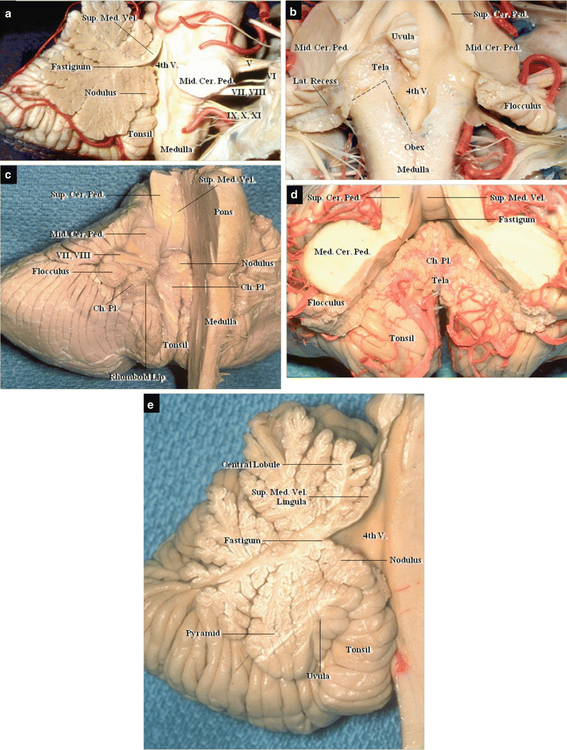
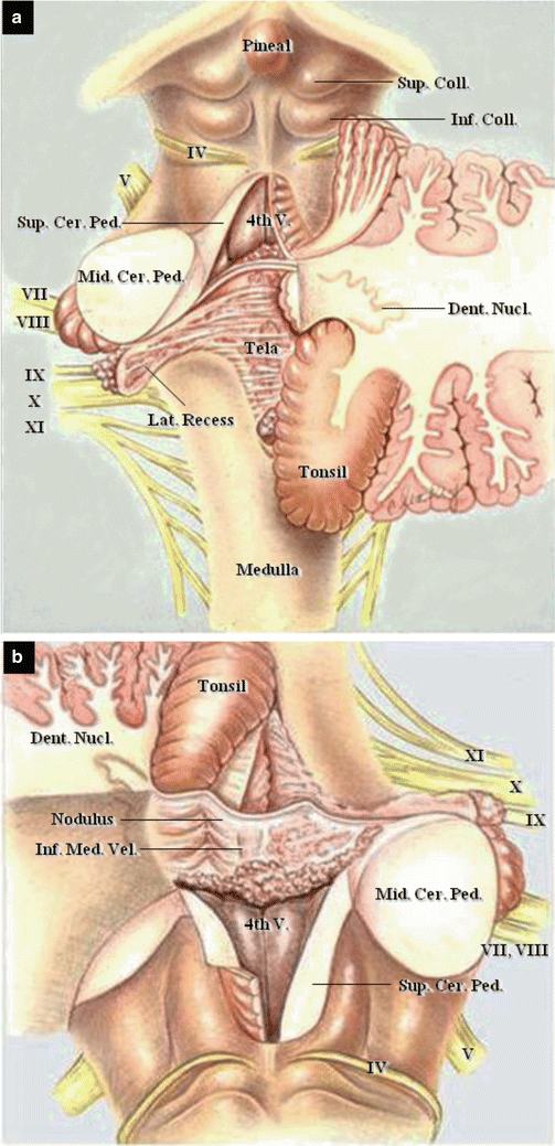
Fig. 2.1
Basic structure of the fourth ventricle. (a) Removal of the right cerebellar hemisphere, showing the fourth ventricle. Right superolateral view. (b) The brainstem after removal of the cerebellum, showing the floor of the fourth ventricle. Superior view. (c) Removal of the right half of the brainstem, showing the roof of the fourth ventricle. Right inferolateral view. (d) The cerebellum after removal of the brainstem, showing the roof of the fourth ventricle. Inferior view. (e) Midsagittal section of the vermis and brainstem
Fig. 2.2
Color illustrations of neural structures in the posterior cranial fossa (from Matsushima T et al. [14] with permission). (a) Superior view of the fourth ventricle. The cerebellar hemispheres were completely removed on the left side and partially removed on the right. (b) Anterior view of the fourth ventricle
2.3 Three Cerebellar Surfaces and Their Surgical Approaches
The cerebellar surface can be divided on the basis of the opposing structures and surgical approach into the tentorial (superior), petrosal (lateral), and suboccipital (posterior) cerebellar surfaces [14, 15]. The tentorial cerebellar surface faces the cerebellar tentorium and is exposed mainly through the infratentorial supracerebellar approach (Fig. 2.3a). The petrosal cerebellar surface faces the petrosal bone and is often exposed and retracted through the lateral suboccipital approach (Fig. 2.4b). The suboccipital cerebellar surface faces the occipital bone and is exposed through the midline suboccipital approach (Fig. 2.5a). Three cerebellar–brainstem fissures exist below the three cerebellar surfaces: the cerebellomesencephalic, cerebellopontine, and cerebellomedullary fissures [14, 15].
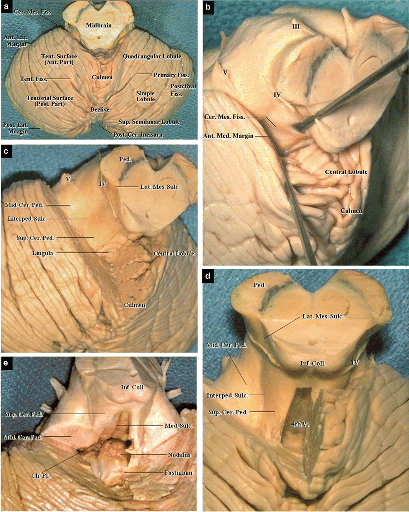
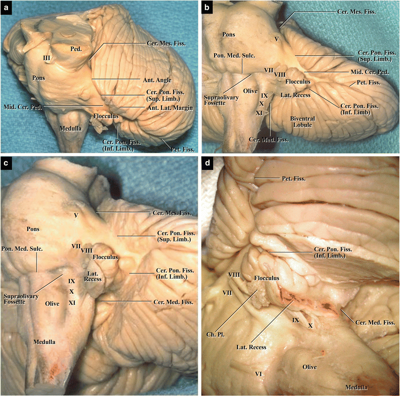
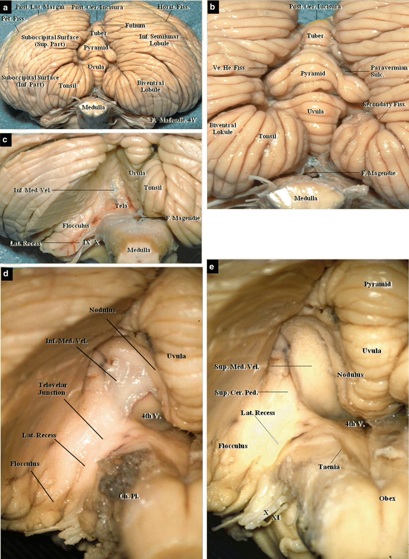
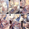
Fig. 2.3
Tentorial (superior) cerebellar surface; step-by-step dissection Superior view. (a) Superior vermis and hemispheres. (b) Left cerebellomesencephalic fissure. (c) Left cerebellomesencephalic fissure after removal of the anterior part of the tentorial cerebellar surface. (d) Anterior view of the fourth ventricle in a specimen with the superior medullary velum removed. (e) Anterior view of the fourth ventricle after removal of the superior cerebellar peduncle
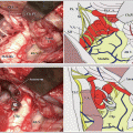
Fig. 2.4
Petrosal (Lateral) cerebellar surface on the left side: step-by-step dissection. (a) Superolateral view. (b) Lateral view. (c) The superior and inferior limbs of the cerebellopontine fissure in a specimen after removal of the lateral margins of the petrosal cerebellar hemisphere. (d) The inferior limb of the cerebellopontine fissure, and the cerebellomedullary fissure, in a specimen with the inferior petrosal cerebellar hemisphere removed
Fig. 2.5
Suboccipital (posterior) cerebellar surface, step-by-step dissection Posterior view. (a) Inferior vermis and hemispheres. (b) High magnification image of the posterior cerebellar incisura. (c) Left cerebellomedullary fissure after removal of the tonsils and biventral lobule. (d) High magnification image of the left cerebellomedullary fissure after reflection of the tela choroidea. (e) High magnification image of the left cerebellomedullary fissure after removal of the tela choroidea and inferior medullary velum
2.4 The Tentorial (Superior) Cerebellar Surface
The tentorial cerebellar surface faces the cerebellar tentorium, sloping anteromedially to posterolaterally in a downward direction (Fig. 2.3a). The superior vermis forms the medial portion: the transition from vermian to hemispheric surface is smooth and lacks the deep fissures evident on the suboccipital cerebellar surface. The tentorial and petrosal cerebellar surfaces are separated by the anterolateral margin. The tentorial and suboccipital cerebellar surfaces are separated by the posterolateral margin. The large fissure, which is situated near the posterolateral margin of the tentorial cerebellar surface, is called the postclival fissure.
The tentorial cerebellar surface is exposed through the infratentorial supracerebellar approach and leads to the tentorial edge region. The cranial nerve (CN) V, Meckel’s cave, and lateral mesencephalic region are also exposed through the lateral infratentorial supracerebellar approach. A deep cerebellar–brainstem fissure, called the cerebellomesencephalic fissure, exists beneath this surface (Fig. 2.3b, c). It is formed by the midbrain and the folded superior vermis of the cerebellum. The roof of the fissure is formed by the culmen and central lobule; the fissure is exposed when the culmen is retracted or resected. The floor of the fissure is formed by the superior medullary velum covered by the lingula medially and the superior cerebellar peduncle laterally (Fig. 2.3c). The interpeduncular sulcus exists lateral to the superior cerebellar peduncle, forming a border between the superior and middle cerebellar peduncles. It is superiorly continuous with the lateral mesencephalic sulcus in the lateral midbrain (Fig. 2.3d). The sulcus, which forms the posterior border of the cerebral peduncle, is a recognized anatomical landmark, indicating the zone of safe approach to cavernous malformations of the midbrain. When the superior medullary velum or superior cerebellar peduncle is incised, the fourth ventricle is exposed from the superior side, thus revealing the nodulus (Fig. 2.3d, e).
2.5 The Petrosal (Lateral) Cerebellar Surface
The petrosal cerebellar surface faces the posterior cranial fossa surface of the petrous temporal bone (Fig. 2.4a, b). Central to the petrosal cerebellar surface is a large fissure, called the petrosal fissure (horizontal fissure), the floor of which is formed by the middle cerebellar peduncle (Fig. 2.4b, c
 The Veins of the Posterior Cranial Fossa: Nomenclature
 The Cerebellopontine Angle: Basic Structures and the “Rules of Three”
 The Temporal Bone: Basic Anatomy and Approaches to Internal Auditory Canal
 Microvascular Decompression for Glossopharyngeal Neuralgia: Surgical Approaches Depending on the Offending Artery
 Microsurgical Anatomy of and Surgical Approaches to the Jugular Foramen
 Microsurgical Anatomy of the Cerebellomedullary Fissure and Variations of the Transcerebellomedullary Fissure Approach


Related posts:
The Veins of the Posterior Cranial Fossa: Nomenclature
The Cerebellopontine Angle: Basic Structures and the “Rules of Three”
The Temporal Bone: Basic Anatomy and Approaches to Internal Auditory Canal
Microvascular Decompression for Glossopharyngeal Neuralgia: Surgical Approaches Depending on the Offending Artery
Microsurgical Anatomy of and Surgical Approaches to the Jugular Foramen
Microsurgical Anatomy of the Cerebellomedullary Fissure and Variations of the Transcerebellomedullary Fissure Approach
Stay updated, free articles. Join our Telegram channel
Full access? Get Clinical Tree
