(1)
Wound Healing Unit, Department of Surgery, Montpellier University, Montpellier, France
8.1 Introduction
The structure of a premature newborn’s skin is very different from that of the adult.
In the adult, the stratum corneum is composed of about 20 layers and has an important protective function. However, in the premature baby, this stratum is non-existent or minimal (0–3 layers). The thinness of these external layers is one of the principal sources of vulnerability (see Fig. 8.1).
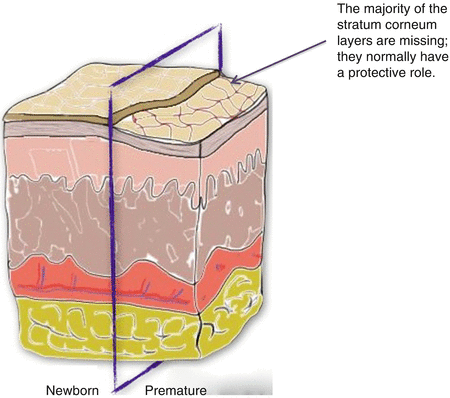
Fig. 8.1
Comparison of normal newborn and premature skin. The stratum corneum is absent or very underdeveloped in the premature skin
The stratum corneum normally protects the body from toxins and infections, enables thermoregulation and controls transepidermal water loss.
In the premature newborn, in addition to the thinness of the external layers, there is also a lack of collagen and elastin in the superficial dermal layers. This considerably increases the risk of pressure ulcers, with spontaneous oedema increasing the cutaneous ischaemia.
In the hospital environment, the incidence of neonatal pressure ulcers (NPUs) is significant, affecting one in four babies in the neonatal intensive care unit (NICU) [1, 2].
Four per cent of babies who have been treated in a NICU are left with a scar [3]. In the majority of cases, these skin lesions are minor [4].
The significant incidence of NPUs in the NICU can be explained by the following risk factors [5]:
The immaturity of the skin
The restriction of the baby’s voluntary movements (e.g. due to a central venous line, mechanical ventilation, etc.)
The hot and humid environment
The use or not of invasive ventilation
This chapter describes the main sources of pressure ulcers in the newborn and premature baby, their characteristic features, the therapeutic principles, and the potential sequelae.
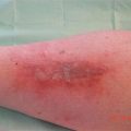
8.2 Risk Assessment Scales
8.3 Principles of Treatment
8.3.1 Topic Treatment
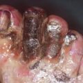
For Stage 2 pressure ulcers, the use of hydrocolloids seems to us to be appropriate in the majority of cases.
For Stage 3 and 4 pressure ulcers, treatment is very similar to that of the adult, but the possibilities available in terms of wound cleaning are often reduced as the baby cannot be moved.
Throughout the liquefactive necrotic stage, the use of silver sulphadiazine as an alternative to hydrogels seems to us to be a possibility. The dressings need to be substantial, thus allowing for additional discharge.
8.3.2 Surgical Treatment
It is reserved for severe cases. It uses the classic reconstruction ladder (skin graft, local and distant flaps). Taking account of the healing capacity of the newborn, the flap realization is often performed in a second step during infant growth.
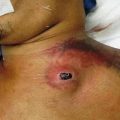
8.4 Main Areas Affected
1.


The nose
The nose is the main area affected, representing half of all NPUs [5]. This has been seen since the 1980s, with the development of nasal continuous positive airway pressure (NCPAP).
According to different studies, nasal lesion incidence is between 20 and 60 % in premature treated with NCPAP [7, 8]. The majority of occurrence is due to the technique being incorrectly used and/or poor monitoring of skin tolerance [9].
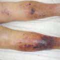
The pressure ulcer most often involves the philtrum, the tip of the nose, the soft triangle, or the nasal septum.
Related posts:


Stay updated, free articles. Join our Telegram channel
Full access? Get Clinical Tree
