Fig. 23.1
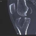
(a, b) The hip center is acquired through pivoting the limb
Surgical Technique
The surgical technique recommended by the authors is a non-anatomical, double-bundle ACL reconstruction, involving fresh-frozen, nonirradiated Achilles tendon allograft with soft tissue fixation [19]. The navigation system (BLU-IGS, Orthokey LLC, Lewes, DE, USA) consists of a commercial optoelectronic localizer and custom acquisition software for kinematic analysis. This is used to intra-operatively measure joint laxity, graft elongations, isometry maps, and previous tunnel placement [20–23] (Fig. 23.2).
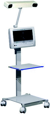
Fig. 23.2
The navigation system (BLU-IGS, Orthokey LLC, Lewes, DE, USA) consists of a commercial optoelectronic localizer and a customable acquisition software for kinematic analysis
The surgical technique involves standard knee arthroscopy with three portals: a medial supra-patellar for water inflow, an antero-lateral for the arthroscope, and an antero-medial for instruments. Menisci and articular cartilage are evaluated and, when necessary, treated. The residual graft from the previous surgery is carefully debrided.
A small 1.5–2 cm skin incision is made over the tibia, just medial to the tibial tubercle. Then, in order to track bone movement during kinematic tests, two reference arrays are fixed, respectively, on the femur and tibia, with 3 mm surgical wires. The tibial reference array is fixed in the approach for tibial tunnel preparation and oriented distally with respect to the knee. The femoral array is inserted above the end of the lateral condyle, distally oriented with respect to the knee (Fig. 23.3). After fixing the femoral and tibial trackers, the surgeon performs an anatomical registration phase, through the percutaneous acquisition of anatomical landmarks: particularly the hip center (identified through pivoting) (Fig. 23.1a, b), femoral epicondyles, tibial malleoli and tibial plateau extremities. Furthermore, other points can be acquired arthroscopically in order to have a complete view of joint characteristics and surgical reconstruction: joint line, tibial plateau centers, and the internal and external exit holes of the previous tunnels. At this point, the system can already determine the placement of the previous tunnels and check for their isometry.
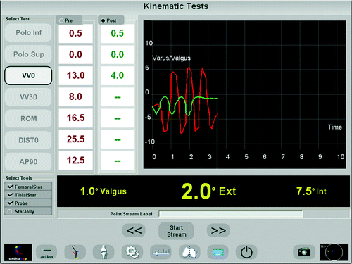
Fig. 23.3
Intraoperative setup
Before performing the ACL revision, a series of kinematic tests is performed in order to evaluate joint kinematics and laxities [20–24]. For revision ACL reconstruction, the tests are:
1.
Passive range of motion (PROM).
2.
Varus-valgus stress test (VV) at 0° (full leg extension) and 30° of flexion.
3.
Internal-external rotation test (IE) at 30 and 90° of flexion.
4.
Antero-posterior stress test (AP) at 30 and 90° of flexion.
5.
Pivot-shift (PS) test.
The 6 degrees of freedom (DoF) of the knee joint are computed from the relative motion of the tibial frame with respect to the femoral one, to decompose instantaneous rotations and displacements. Knee laxities are also defined for each performed test.
The tibial and femoral tunnels are prepared right after this preoperative evaluation. The authors recommend the creation of just one tibial tunnel where both bundles of the graft are passed to avoid additional damage to the bone stock, while achieving a better press-fit of the two bundles. On the femoral side, one tunnel is required for the reconstruction of the postero-lateral bundle of the ACL, while the antero-medial bundle is reconstructed through an over-the-top passage of the graft, avoiding additional tunnel creation.
According to the technique [19], the knee is flexed to about 135° and a guide pin is drilled through the tibia at an angle of 55° to position it in the postero-medial part of the ACL footprint. For the femoral tunnel, the knee is repositioned a little less than 90° of flexion and the guide pin is inserted through the antero-medial portal on the medial wall of the lateral condyle, approximately 5 mm anterior to the over-the-top position. Then the knee is flexed to around 130°, and the guide pin is advanced until it passes the femoral cortex, just above the end of the lateral femoral condyle. The internal and external exit point of the guide pins on both tibia and femur are collected by the system (Fig. 23.4). Digitizing the diameter of the reamer, chosen by the surgeon to drill the tunnels, it is possible to check the isometry of the new tunnels and search for any possible overlapping with the previous ones.
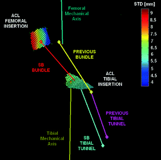
Fig. 23.4
Maps of isometry for ACL tibial and femoral insertions, with the corresponding previous graft placement and the position of the new postero-lateral bundle
If the placement of the tunnels is good, both bundles of the graft are passed through the tibial tunnel and then, while the antero-medial bundle is brought in the over-the-top position through the posterior capsule, the postero-lateral one is passed throughout the femoral tunnel.
Finally, after the fixation of the graft, the kinematic evaluation is repeated to check real-time the efficacy of the reconstruction and to search for any possible associated secondary laxity (Fig. 23.5).

Fig. 23.5
Typical results of laxity reduction after ACL revision
Kinematic evaluation of intra-operative data is performed off-line with dedicated software [25, 26] designed for the study of diarthrodial joints. The 3D coordinates of the anatomical landmarks are used to compute the bone reference frames [27, 28]. Motion data, recorded during kinematic tests, are elaborated using Euler decomposition [20]. VV laxities are calculated along the axis identifying anterior-posterior direction on tibia. IE laxities are calculated along tibial mechanical axis. AP translations are calculated as the 3D displacement of the tibia reference frame centered in the most posterior point of ACL tibial insertion.
Discussion
ACL revision surgery is a challenging operation and its results remain poor when compared with primary procedures. Many studies comparing navigated and conventional ACL reconstruction have proven that CAOS produces better tunnel placement, eliminates impingement, and reduces anisometry in cases of ACL reconstruction. This improvement is especially effective with relatively inexperienced surgeons, an important fact because more than 80 % of ACL surgery is performed by this group [17].
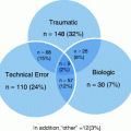
While all these advantages of CAOS are available for primary ACL reconstruction, they are especially important for ACL revision surgery [18]. Indeed, in revision surgery CAOS might be effective to improve its outcome and to detect causes for failure of the primary reconstruction. ACL reconstruction fails due to problems in technique, graft incorporation, and reinjury [13, 14, 17]. The majority of cases relate to technical errors; mal-positioning of the tibial and femoral tunnels forming the bulk of the problem. Almost all of the technical errors of ACL reconstruction are correctable using CAOS. Mal-positioned tunnels (the primary reason for graft failure), graft impingement, and anisometry are the three factors that can be avoided by navigation in revision surgery [13, 14, 17


Related posts:

Stay updated, free articles. Join our Telegram channel
Full access? Get Clinical Tree
