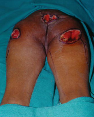
Fig. 12.1
Anteroposterior (AP) x-ray of the pelvis of a patient with multiple pressure ulcers showing destruction of both ischial bones secondary to chronic ulceration and infection
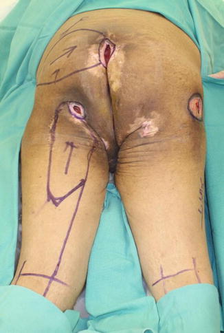
Fig. 12.2
Operative photograph of the patient in the prone position with multiple pressure ulcers, stage IV, sacral, left ischial, and right trochanteric ulcers showing the marking of the design of the flaps to close these ulcers
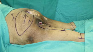
Fig. 12.3
Operative photograph, left lateral view, of the same patient showing the marking of the left gluteus maximus sliding island flap
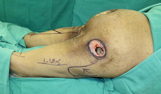
Fig. 12.4
Operative photograph, right lateral view, of the patient showing the design of the rotation tensor fascia lata flap
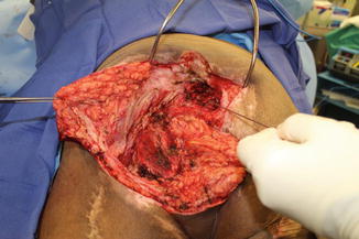
Fig. 12.5
Operative photograph showing the excision of the left ischial ulcer, the left ischium shaved, and excision of the coccygeal bone. There was communication between the ischial and the coccygeal ulcer and, for this reason, the gluteus maximus flap was utilized as a rotation flap
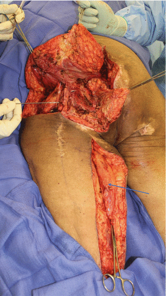
Fig. 12.6
Operative photograph showing the complete dissection of the gluteus maximus to expose the entire area. The metal wire indicates the position of the sciatic nerve. The gracilis muscle was dissected and exposed. Arrow indicates the position of the gracilis muscle
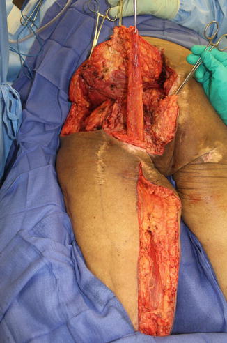
Fig. 12.7
Operative photograph showing the gracilis muscle tunneled to the left ischiococcygeal area
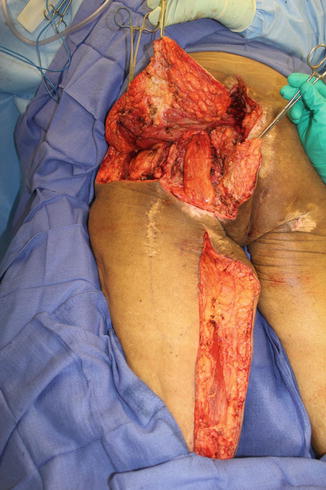
Fig. 12.8
Operative photograph showing the position of the gracilis muscle, which covers the ischium and the coccygeal bone in the same time
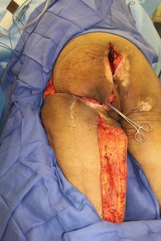
Fig. 12.9
Operative photograph showing the medial rotation of the gluteus maximus flap to cover the defect
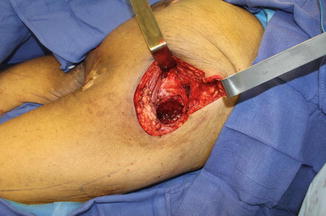
Fig. 12.10
Operative photograph showing the excision of the right posterior trochanteric ulcer and bursa with shaving of the right trochanteric bone
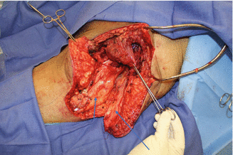
Fig. 12.11
Operative photograph showing the dissection of the lower portion of the gluteus muscle to be the first layer covering the trochanteric bone. The right tensor fascia lata (TFL) flap was raised. Arrow indicates the TFL fascia. Short arrow indicates the vastus laterals muscle
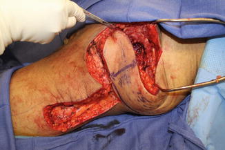
Fig. 12.12
Operative photograph showing the dissected tensor fascia lata (TFL) flap with rotation to cover the entire defect
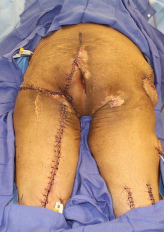
Fig. 12.13
Operative photograph showing the complete closure of the left gluteal flap and left gracilis donor site. On the right side, the two incisions were used to release the hamstring muscles to treat contractures. The right side shows the sutured TFL flap
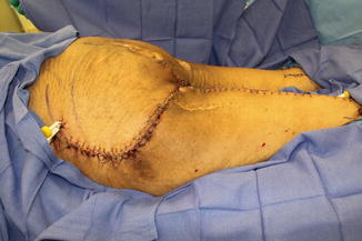
Fig. 12.14
Operative photograph, left lateral view, showing the closure of the left gluteal flap
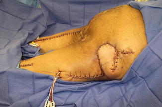
Fig. 12.15
Operative photograph, right lateral view, showing the closure of the right tensor fascia lata (TFL) flap and the donor site of the flap
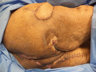
Fig. 12.16
Photograph, 6 weeks post-operative, showing healed right tensor fascia lata (TFL) flap
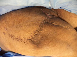
Fig. 12.17
Photograph, 6 weeks post-operative, showing healed left gluteus maximus flap
