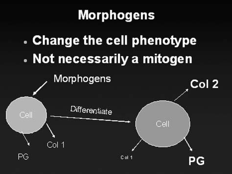
48 Biological treatment of disk degeneration encompasses a large diversity of different therapeutic approaches.1–14 In broad categories, the therapeutic strategies under investigation involve the use of cells, matrix, or molecules. This chapter focuses on the molecular therapy approaches to treat disk degeneration. The term therapeutic molecule will be used as a broader way of encompassing all molecules that may be therapeutic but may not be classic “growth factors.” This terminology is important because growth factors are defined by their effect on mitosis; however, effective therapeutic molecules may have therapeutic activity unrelated to cell replication. The therapeutic molecules can be categorized as anticatabolics, mitogens, chondrogenic morphogens, or intracellular regulatory molecules (Table 48–1). This chapter reviews key literature and defines each of these categories.
Molecular Therapy of the Intervertebral Disk
Category | Molecule |
Anticatabolic | TIMP-1 |
| TIMP-2 |
Mitogens | IGF-1 |
| PDGF |
| EGF |
| FGF |
Morphogen | TGF-b |
| BMP-2 |
| BMP-7 (OP-1) |
| BMP-13 (GDF-6 aka CDMP-2) |
| GDF-5 (CDMP-1) |
| Link N |
Intracellular regulators | SMADs |
| Sox9 |
| LMP-1 |
BMP, bone morphogenetic protein; CDMP, cartilage-derived morphogenetic protein; EGF, epidermal growth factor; FGF, fibroblast growth factor; GDF, growth and differentiation factors; IGF, insulin-like growth factor; LMP, LIM Mineralization protein-1; OP-1, osteogenic protein-1; PDGF, platelet-derived growth factor; SMAD, T/C; TGF-b, transforming growth factor-beta; TIMP, tissue inhibitors of matrix metalloproteinases.
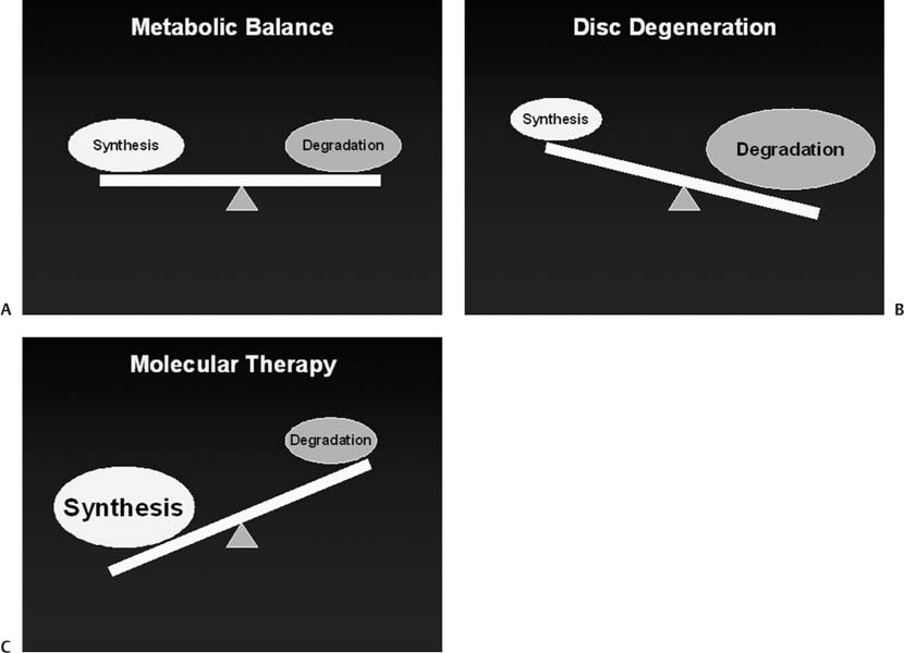
To better understand molecular therapy strategies, it is important to understand the hallmarks of disk degeneration. These include loss of proteoglycan, water, and collagen II in the disk matrix in the nucleus pulposus. There are qualitative changes in the matrix that are less well defined, including loss of the higher molecular weight proteoglycans and other changes that are more difficult to quantify (collagen cross-linking, organization of the proteoglycan, etc.). An important change seems to be the loss of the differentiated chondrocyte phenotype from the nucleus pulposus into a more fibrotic phenotype. Changes in the annulus fibrosus include disorganization of the annular lamella layers and physical defects in the collagenous matrix. Typically, these matrix changes take many years to become apparent and are most likely a result of an imbalance between synthesis and degradation (Fig. 48–1). The goal of molecular therapy is to prevent or reverse these changes in the disk extracellular matrix by altering the balance of degradation to synthesis in favor of synthesis.
Anticatabolics
Because matrix loss is a balance between matrix synthesis versus degradation, it is possible to increase disk matrix by increasing synthesis or by decreasing degradation. One approach is to prevent matrix loss by inhibiting the degradative enzymes. There are many different catabolic enzymes present in normal disk matrix, but the matrix metalloproteinases (MMPs) make up a particularly important group of catabolic enzymes.15 MMPs play an important role in the normal turnover of matrix molecules and are probably important in disk degeneration.16 Degenerated disks have elevated levels of MMPs, providing circumstantial evidence to support this hypothesis. Within the matrix, MMP activity is normally inhibited by tissue inhibitors of MMPs (TIMPs).15,17 Wallach et al tested whether one of these anticatabolic molecules, TIMP-1, could increase the accumulation of matrix proteoglycans in in vitro experiments using an adenoviral gene therapy approach.18 Wallach et al found that, indeed, TIMP-1 expression in disk cells increased accumulation and also increased the “measured synthesis rate” of proteoglycans. Another promising molecule is CPA-926 an esculetin prodrug that has a better pharmacokinetic profile than esculetin itself.19 CPA-926 has been shown to be anti-inflammatory and antitumorigenic and to prevent degeneration in an osteoarthritic model of cartilage destruction.19 Okuma et al showed that oral administration of CPA-926 can prevent or delay the onset of disk height loss and histological evidence of disk degeneration in an annulotomy model of disk degeneration in the rabbit.19
Figure 48–1 Disk matrix metabolism: balance of synthesis and degradation. (A) In the homeostatic state, the disk undergoes matrix synthesis and degradation in a balanced manner. (B) As the disk matrix undergoes turnover over the course of an individual’s lifetime, any small imbalance between synthesis and degradation can lead to significant changes in overall disk matrix content. (C) One of the major goals of molecular therapy of the disk involves modulating this metabolic balance to the more favorable anabolic state. This can be accomplished by increasing synthesis or by decreasing catabolism.
Along with the balance of synthesis and degradation, the rate of disk matrix metabolism may also be important. For example, the overall rate of disk metabolism in young disks may be much higher than in old disks. This may lead to qualitative changes in disk matrix such as the composition of degraded aggrecan versus newly synthesized aggrecan molecules. The overall metabolic rate may also dictate the nutritional requirements of the intradiskal cells. Cytokines such as interleukin (IL)-1 and tumor necrosis factor-alpha (TNF-a) may have critical roles in disk metabolism. Therefore, molecules such as IL-1Ra and infliximab, which can block IL-1 and TNF-a, respectively, may be useful.20–22 Further research into anticatabolic molecules may yield important results.
Mitogens
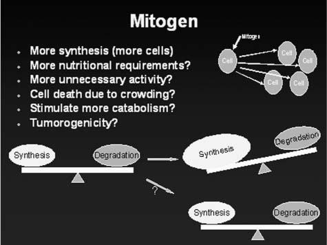
Mitogens are defined by the propensity of the molecule to increase the rate of mitosis. These molecules constitute the true growth factors, and for purposes of this review, we will differentiate mitogenic growth factors from the set of molecules that are highly chondrogenic (Fig. 48–2). These mitogenic molecules include insulin-like growth factor-1 (IGF-1), epidermal growth factor (EGF), and fibroblast growth factor (FGF).23 Thompson et al23 demonstrated in vitro experiments with mature canine disk cells that mitogenic molecules can increase the rate of mitosis and proteoglycan synthesis rates to various degrees depending on which region of the disk the cells were derived from. In general, EGF performed better than the other mitogens.23 IGF-1 levels decrease in an age-dependent manner in rat disks.24 Researchers have speculated that by restoring the IGF-1 in aging disks that perhaps matrix synthesis may be increased. In vivo experiments with growth factors using a mouse tail disk compression model for degeneration by Walsh et al25 produced results that were consistent with in vitro experiments by Thompson et al.23 IGF-1 had mild effects (especially in the inner annulus) and FGF had no effect. In a related but different potential mechanism of therapy, some growth factors may protect disk cells from death by apoptosis. Gruber et al26 showed that disk cells in vitro placed in low serum conditions underwent apoptosis; however, this was prevented to a certain degree by the addition of insulinlike growth factor (IGF-1) or platelet-derived growth factor (PDGF). Although IGF-1 has some anabolic effects, it may also have an effect on catabolism because IGF-1 was shown to decrease the levels of active TIMP-2 in tissue culture experiments, indicating a complex effect on disk matrix metabolism by IGF-1.
Figure 48–2 Mitogenic molecules are the true growth factors. They increase cell number without necessarily enhancing cell differentiation. The increase in cell number leads to increase in matrix synthesis. However, this may also increase the nutritional requirement in the disk due to higher overhead of keeping the cell alive and perhaps increase the overall catabolism as well as synthesis. Furthermore, very high cell density can lead to cell death with decreases in disk nutrition. Finally, mitogens may have a higher likelihood of causing tumors as opposed to morphogens, which increase cell differentiation.
Figure 48–3
Stay updated, free articles. Join our Telegram channel

Full access? Get Clinical Tree


 Anticatabolics
 Mitogens
 Morphogens
 Intracellular Regulators
 Summary


