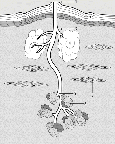
Fig. 11.1
Diagram of types of nipple: (a) everted erect (normal); (b) everted flat (normal); (c) inverted umbilicated (normal congenital); (d) true inverted (pathologic usually benign); (e) partially retracted or deviated (pathologic, both benign and malignant); (f) fully retracted (pathologic, usually malignant)
Everted nipple is the common (normal) type, where nipple is held erect by a cylindrical column of small smooth muscles. Even if its projection is low or flat, when stimulated by touch or cold, it becomes erect.
Inverted nipple appears to be indented in the areola, and in congenital cases, this aspect is due a failure of the underlying mesenchymal tissue to proliferate and project the nipple papilla outward. The nipple may be pulled out with gentle squeezing of the areolar skin, and the projection could be maintained for some minutes then the nipple reverts to an inverted state. Inverted nipples may be:
Congenital (about 3–10 % of normal types, prevalence may change with age, pregnancies, and lactation) that may be umbilicated (more than 90 % of congenital forms, easy to pull out) or invaginated (3–10 % of congenital forms, more difficult to pull out)
Acquired when a forceful manipulation is required to pull the nipple out, and inversion recurs quickly. This type of nipple inversion may occur because the nipples are stuck into scar tissue of the periductal mastitis (commonly presents as a slitlike inversion, Fig. 11.2) or into fibrosis due to aging or as late duct ectasia phenomenon (commonly presents as a circular inversion).
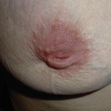
Fig. 11.2
Slitlike nipple inversion, symmetrical and central, involving the nipple but not the areola
Retracted nipples, unlike inverted nipples, will not come back out when stimulated. The nipple is strongly deviated or buried below the level of the skin. Despite maximal manipulation, the nipple cannot be pulled out. Nipple retraction may be partial (deviated nipple), when a single duct is involved in a pathological process as periductal mastitis or invasive/ non-invasive ductal carcinoma (Fig. 11.3), or full, as result of severe fibrosis or pathological causes as chronic inflammatory diseases and carcinoma.
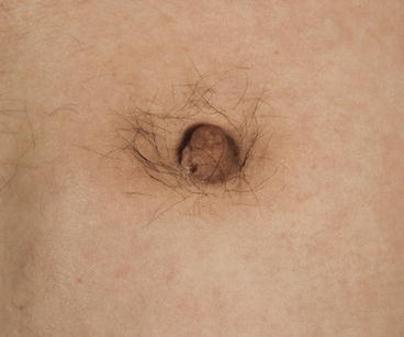

Fig. 11.3
Nipple retraction, due to underlying BC in a man
A synopsis of nipple types is showed in Table 11.1 that includes also a grading system for nipple inversion based upon how difficult it is to pull the nipple out manually and how well projection is subsequently maintained. The majority of patient with nipple inversion have grade II inversion.
Table 11.1
Classification of nipple types
Definition | Types | Causes and notes | Grade |
|---|---|---|---|
Everted | Erect | Normal common | Grade 0 |
Flat | Flat type became protractile when compressed or stimulated | ||
Inverted | Umbilicated | Normal congenital | Grade I – the nipple is pulled out with gentle squeezing of the areolar skin. Nipple projection is well maintained for several minutes, but then the nipple reverts to an inverted state |
>90 % of congenital easy to pull out | Due to a failure of the underlying mesenchymal tissue to proliferate and project the nipple papilla outward | ||
Invaginated | 3–10 % of normal types, prevalence may change with age, pregnancies, and lactation | ||
5–10 % of congenital difficult to pull out | |||
True inverted | Acquired | Grade II – forceful manipulation is required to pull the nipple out, and inversion recurs quickly | |
Slitlike inversion (due to periductal mastitis) | |||
Circular inversion (fibrosis due to aging or as late duct ectasia phenomenon) | |||
Retracted | Partially retracted (deviated) | Acquired | Grade III – the nipple is strongly deviated or buried below the level of the skin. Despite maximal manipulation, the nipple cannot be pulled out |
Fully retracted | Deviated (scarring of a single duct due to periductal mastitis, prior surgery or breast cancer) | ||
Fully retracted (severe fibrosis due to aging, or duct ectasia phenomenon, pathological processes as chronic inflammatory diseases and BC) |
SURGICAL CORRECTION OF BENIGN NIPPLE INVERSION – For many women, inverted or non-protractile nipples can be a source of aesthetic and functional concern, leading to self-consciousness and psychological distress.
The goal of surgical correction is to restore projection while maintaining the ductal anatomy as much as possible. For this purpose, the simplest nonsurgical technique is a plain device that can be used by women of any age to pull out inverted or non-protractile nipples. Using suction to gently stretch the ducts over time, it can achieve a permanent correction between 1 and 3 months of continuous use, for 8 h/ day.
The simplest surgical technique is a purse-string suture, which is placed around the neck of the nipple through a periareolar incision. This technique tightens the neck of the nipple and works well for those with less severe cases of nipple inversion.
Selective division of ducts is another technique that makes a blunt dissection through vertical spreading of fibrous tissue parallel to the lactiferous ducts through an inferior periareolar incision. Placing the nipple on traction for 2–5 days with a stent completes the repair. More severe cases of nipple inversion are treated with triangular skin flaps, which can be mobilised to add bulk to the base of the nipple and, when closed, tighten the neck of the nipple [2].
If ducts may be carved, percutaneous release of nipple inversion can be accomplished with a simple minimally invasive technique using a needle tip for lysis of retracted ducts. For this technique, an 18-gauge needle tip is inserted at the 6 o’clock position and used to lyse fibrous tissue and tethered glands until satisfactory nipple projection has been achieved. Following this, a monofilament purse-string suture is placed, starting at the entry site, with entry and exit through the same stitch point every 3–5 mm around the nipple base. The suture is then tied under moderate tension. In a series of 58 inverted nipples in 31 patients, there were 13 recurrences, of which 11 were successfully treated with a second purse-string suture and two required a third procedure [3].
11.1.2 Nipple Adenoma
Nipple adenoma is an uncommon and distinctive variant of intraductal papilloma, a benign proliferative lesion restricted to the nipple [4]. Alternative terms for this entity include mainly florid papillomatosis but also erosive adenomatosis, syringomatous adenoma, superficial papillary adenomatosis, and papillary adenoma. The spectrum of clinicopathologic features related to its unique location and heterogenous histopathology makes nipple adenoma a unique entity.
Nipple adenoma may occur at any age even though is most commonly seen in the middle aged to elderly. Asymptomatic nipple adenoma is found in 1–16 % of breast specimens obtained for carcinoma. It usually presents as a papilliferous lesion of the nipple, often with ulceration, which may be concealed under a crust. The condition is sometimes painful, the commonest descriptions being burning or itching.
On examination, the whole nipple may be indurated, has an expanded appearance, and may be ulcerated. The main differential diagnoses are prolapsing intraductal papilloma, Paget’s disease of the nipple, factitial disease, and eczema.
The differential diagnosis from Paget’s disease can only be made with certainty by biopsy, although the characteristic clinical features are different. Nipple adenoma does not extend on to the areola like established Paget’s disease, and it has the appearance of a deeper lesion eroding through the nipple. Early Paget’s disease is more superficial in appearance.
Nipple adenoma erodes through the nipple duct (Fig. 11.4), while an intraductal papilloma, if (rarely) superficial and prolapsing through the duct, tends to expand the nipple rather than erode it (Fig. 11.5). If a papilloma prolapses through the opening of a duct, the nipple remains normal and never becomes ulcerated.
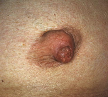
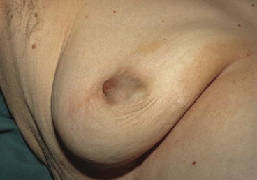

Fig. 11.4
Nipple adenoma, involving only the nipple area

Fig. 11.5
Large central papilloma deforming the areola with minimal involvement of the nipple
A comparison among clinical features of nipple adenoma, Paget’s disease, and prolapsing intraductal papilloma is showed in Table 11.2.
Table 11.2
Compared clinical features of prolapsing intraductal papilloma, nipple adenoma, and Paget’s disease
Prolapsing intraductal papilloma | Nipple adenoma | Paget’s disease |
|---|---|---|
Does not extend on the areola | Does not extend on the areola | Areola is involved in most cases |
Tends to expand the nipple rather than erode it | Erodes through the nipple as a deeper lesion | Is superficial in appearance |
Never ulcerated | Sometimes ulcerated | Crusting but never ulcerated |
Painless or slightly burning | Painful, sometimes burning or itching | Slightly itching |
Benign at low risk (if no atypia) | Benign at low risk | Preinvasive cancer |
Nipple adenoma is not itself regarded as premalignant, although, in about 5 % of cases, it has been found associated with BC. This incidence of associated BC needs for careful assessment of both breasts. Another association of nipple adenoma is with intraductal papilloma that has also been reported to occur more commonly than would be expected by chance. Nipple adenoma is adequately treated by local excision of the affected part of the nipple, and in most cases there is no need to remove the whole nipple. This assertion is anyway still controversial.
11.1.3 Observations About Montgomery’s Glands
Three types of glands are found in the areolar skin:
Get Clinical Tree app for offline access

Apocrine sweat glands, which are a normal finding, with no clinical implication
Modified sebaceous glands (Montgomery’s glands) which are similar to sebaceous glands elsewhere, except that they are associated with a lactiferous duct extending from a more deeply placed rudimentary mammary gland (Fig. 11.6)

