(1)
Department of Neurosurgery Faculty of Medicine, Saga University, Saga, Japan
Keywords
Trigeminal neuralgiaMicrovascular decompressionInfratentorial lateral supracerebellar approachTentorial stitched sling retraction method11.1 Introduction
Microvascular decompression (MVD) using the lateral suboccipital approach (LSA) has become the treatment of choice for trigeminal neuralgia (TN) since Jannetta’s original description using a surgical microscope [3, 4]. The goal of this functional surgery is to prevent postoperative complications such as hearing loss and to achieve complete pain relief. Recurrence of pain remains a problem after MVD; therefore, every effort should be made to prevent it [5–7, 14].
Two slightly different types of LSA have been used during MVD for TN: the infratentorial lateral supracerebellar approach [7, 9, 10, 12] and the combined transhorizontal-supracerebellar approach [1]. These two approaches, which partly overlap each other, are suitable for surgeries around the Meckel’s cave, particularly decompression of the trigeminal nerve [cranial nerve (CN) V]. Because the combined approach is already described in Chap. 9, “The Retrosigmoid Lateral Suboccipital Approach: Basic Approach and Variations,” this chapter presents the infratentorial lateral supracerebellar approach and the tentorial stitched sling retraction method [6–9].
In more than 80 % cases of TN, the offending vessel is the superior cerebellar artery (SCA). Therefore, the supracerebellar approach from the medial side and the method to displace SCA superomedially and anchoring it are extremely reasonable for achieving decompression and preventing its recurrence. After introducing the tentorial stitched sling retraction method, we experienced only three cases (5 %) of recurrence among 63 continuous surgical cases at Saga University Hospital, Saga, Japan between 2007 and 2013; moreover, these cases did not require reoperation because the pain was mild.
Decompression of an elongated tortuous vertebral artery (VA) is also described at the end of this chapter because it slightly differs from that of an offending SCA or anterior inferior cerebellar artery (AICA).
11.2 History of the Development of Microvascular Decompression for Trigeminal Neuralgia
During the developmental phases of MVD, various surgical approaches and decompression techniques were employed as MVD [3–5, 15]. When MVD for TN was first used in Japan, approximately 30 years ago, it was performed using an approach that was almost identical to that used for acoustic tumors, which used the petrosal (lateral) cerebellar surface alone. In 1987, we reported the usefulness of the infratentorial lateral supracerebellar approach for TN, emphasizing utilization of the tentorial (superior) cerebellar surface [9, 10]. In 2000, Fujimaki T et al. reported the combined approach in which both the petrosal and tentorial cerebellar surfaces were used during MVD for TN [1]. From the viewpoint of safe handling of the superior petrosal veins (Sup. Pet. Vs.), the combined approach may be better when performed completely because the offending vessels are decompressed after complete dissection of the arachnoid membrane around all tributaries of the Sup. Pet. V. However, when the combined approach is insufficiently performed and the tentorial cerebellar surface is insufficiently exposed, the offending SCA is decompressed from the posterolateral side on the petrosal cerebellar side. This direction of transposition is unsuitable for shifting the offending SCA medially. The infratentorial lateral supracerebellar approach, conversely, is extremely suitable for medial transposition of SCA [8, 9].
Subsequently, decompression techniques have also been improved. When MVD for TN was initially employed, the interposing method, in which a prosthesis is inserted between an offending vessel and CN V, was generally utilized [4, 14, 15]. However, after experiences of recurrence due to prosthesis adhesion to CN V, interposition was gradually superseded by transposition [5, 14]. Hence, we propose the use of the tentorial stitched sling retraction technique [6–8]. The causes of TN were also clarified as surgical experience increased and are currently considered to include not only offending arteries but also offending veins and/or arachnoiditis [9, 11].
11.3 Merits of the Infratentorial Lateral Supracerebellar Approach
Using the infratentorial lateral supracerebellar approach, surgeons can reach CN V passing on the lateral portion of the tentorial cerebellar surface medial to the vein of the cerebellopontine fissure, the largest common stem of the Sup. Pet. V [7–10]. The approach has the following advantages:
The risks of postoperative complications such as hearing disturbance and facial palsy are reduced because the approach is distant from CNs VII and VIII.
The relationship between the medial surface of CN V and the offending SCA is clearly visible, and medial transposition of the artery is easy.
The tentorial stitched sling retraction technique can be performed easily and safely because the operative field on the tentorial cerebellar surface using this approach is extremely wide.
The Meckel’s cave can be observed from a medial direction, and the suprameatal bulging does not disturb the view through the surgical microscope.
Care must be taken to sufficiently dissect the arachnoid membrane around the Sup. Pet. Vs., particularly the anterolateral marginal vein and the vein of the cerebellopontine fissure.
11.4 Surgical Anatomy for the Infratentorial Lateral Supracerebellar Approach
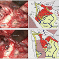
The entry zone of CN V is located just anteroinferior to the anterior angle, which is the most anterior end of the anterolateral margin separating the tentorial and petrosal cerebellar surfaces (Fig. 11.1a). Therefore, CN V can be reached by following this margin, which is situated parallel to the superior petrosal sinus. One or two common stems of the Sup. Pet. V. usually exist between the porus acusticus internus (PAI) and the Meckel’s cave [13] (Fig. 11.1c). The suprameatal bulging, which is a bulge of the petrous bone between PAI and the Meckel’s cave, is sometimes large and can disturb visualization of the Meckel’s cave [16] (Fig. 11.1b). The lateral pontomesencephalic segment of SCA usually courses medial to CN V, bifurcating into the rostral and caudal trunks and forming a downward loop. When the downward loop is long, it compresses the nerve from the superomedial side and becomes the offending vessel for TN [2, 9] (Fig. 11.1d, e). Because perforators originating from SCA are long, SCA can be easily transposed medially (Fig. 11.1e). An upward loop of AICA courses inferolateral to CN V and sometimes compresses it from the inferolateral side [8, 9] (regarding the anatomy of SCA and AICA, refer to Chap. 3, “Three Cerebellar Arteries: Superior Cerebellar Artery, Anterior Inferior Cerebellar Artery, and Posterior Inferior Cerebellar Artery,” and Chap. 10, “Anatomy for Microvascular Decompression Procedures: Relationships between Cranial Nerves and Vessels, Preoperative Images, and Anatomy for the Stitched Sling Retraction Technique”).
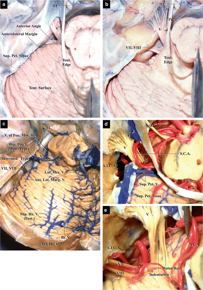
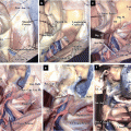
Fig. 11.1
Basic surgical anatomy for the infratentorial lateral supracerebellar approach to cranial nerve V (from Matsushima T [8] with permission). (a) Neural structures. Left tentorial (superior) cerebellar surface. Superior view: the left CN V is situated just in front of the anterior angle. (b) Neural structures. The anterior part of the left tentorial cerebellar surface has been removed and the entry zone of CNs V, VII, and VIII is clearly exposed. (c) Venous structures. Left cerebellopontine angle and tentorial cerebellar surface. Superior view: two petrosal veins exist between the porus acusticus internus and the Meckel’s cave. One bridging vein can be seen in the posteromedial portion of the tentorial cerebellar surface. (d) Arterial structures. The structures surrounding the left CN V, particularly the SCA superior view: the lateral pontomesencephalic segment of SCA compresses CN V from the medial side. (e) Arterial structures. An indentation is clearly visible on CN V when SCA is elevated
11.5 Procedures and Methods for the Infratentorial Lateral Supracerebellar Approach
11.5.1 Positioning
Before positioning, a lumbar drainage is placed in order to exclude the cerebellospinal fluid (CSF). The patient is placed in the lateral park bench position. The head is rotated slightly away from the affected side and the neck is flexed slightly. However, the degree of neck flexion required in patients with TN is less than that required in patients with hemifacial spasm (HFS), which is treated using the infrafloccular approach. The patient’s shoulder is taped and pulled to create a working area wide enough for a surgeon’s arm. However, too strong a retraction of the shoulder may cause brachial plexus injuries (regarding patient positioning, refer to Chap. 9: “The Retrosigmoid Lateral Suboccipital Approach: Basic Approach and Variations”).
11.5.2 Skin Incision and Bony Opening
The posterior end of the incisura mastoidea, which is a palpable bony groove under the skin located just behind the mastoid process, broadly corresponds to the PAI level. Craniotomy for TN should be performed above this level. A gentle S-shaped incision measuring approximately 6–7 cm is made slightly medial to the hairline. The 3–4-cm superior portion of the incision should be made above the incisura mastoidea and the remaining 3 cm should be below it. The upper edge of the skin incision should end approximately 2 cm above the auricle. The occipital artery and nerve are encountered when the nuchal muscles are incised. Although the occipital artery has to be cut, the occipital nerve should be preserved as much as possible.
Related posts:
 The Veins of the Posterior Cranial Fossa: Nomenclature
 The Cerebellopontine Angle: Basic Structures and the “Rules of Three”
 The Temporal Bone: Basic Anatomy and Approaches to Internal Auditory Canal
 Microvascular Decompression for Glossopharyngeal Neuralgia: Surgical Approaches Depending on the Offending Artery
 Microsurgical Anatomy of and Surgical Approaches to the Jugular Foramen
 Microsurgical Anatomy of the Cerebellomedullary Fissure and Variations of the Transcerebellomedullary Fissure Approach
Stay updated, free articles. Join our Telegram channel

Full access? Get Clinical Tree


