(1)
Department of Neurosurgery Faculty of Medicine, Saga University, Saga, Japan
Keywords
Hemifacial spasmMicrovascular DecompressionLateral suboccipital approachInfrafloccular approach12.1 Introduction
We describe the approaches and techniques for microvascular decompression (MVD) for hemifacial spasm (HFS), focusing particularly on the lateral suboccipital infrafloccular approach. The lateral suboccipital infrafloccular approach for HFS differs completely from the approach for trigeminal neuralgia, although both approaches are termed as lateral suboccipital approach (LSA) [4, 8–10, 13–15]. The root exit zone (REZ) of cranial nerve (CN) VII or a nearby area is usually responsible for vascular compression-related HFS; therefore, we recommend the lateral suboccipital infrafloccular approach, in which REZ of CN VII is approached through the space between the origin of CN IX and the flocculus and choroid plexus protruding the foramen of Luschka from the inferior side [4, 7, 10, 13–15]. This approach can be used not only for MVD for HFS but also for other surgeries around the supraolivary fossette such as acoustic schwannoma surgery or pontomedullary tumor surgery.
12.2 Advantages of the Infrafloccular Approach
Several postoperative complications of MVD have been reported [3, 10]. One of the most frequent complications of MVD for HFS is postoperative hearing disturbance due to injury of CN VIII caused by retraction of the petrosal (lateral) cerebellar surface. During cerebellar retraction through the conventional LSA, the flocculus is the biggest obstacle to the view of REZ of CN VII [10, 14, 15, 17]. In the infrafloccular approach, however, the inferior portion of the flocculus and the choroid plexus is gently retracted superiorly and the lateral portion of the pontomedullary sulcus, especially the REZ of CN VII, can be observed through the space between the origin of CN IX and the flocculus. Because the flocculus is retracted in a direction perpendicular to CN VIII, damage to CN VIII can be reduced [10, 14].
12.3 Basic Surgical Anatomy for the Infrafloccular Approach
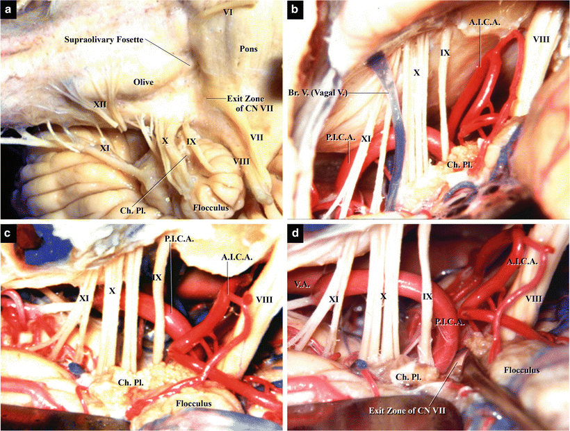
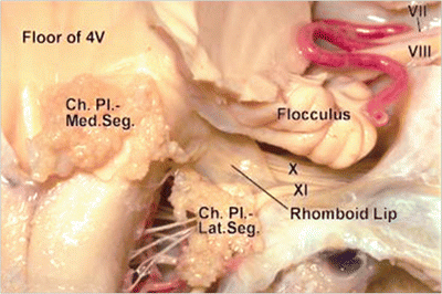
Important anatomical landmarks on the brainstem for cerebellopontine angle (CPA) surgeries include the flocculus and the choroid plexus, which protrudes from the foramen of Luschka (Fig. 12.1a). CNs VII and VIII are located superomedial to the flocculus and the lower CNs are inferior to it. The foramen of Luschka is formed by a membrane of neural tissue called the rhomboid lip (Fig. 12.2a). The choroid plexus and rhomboid lip are situated on the origins of CNs IX and X (Fig. 12.1a). Occasionally, the flocculus and/or rhomboid lip is large and becomes an obstacle (Fig 12.2b, c). REZ of CN VII is situated immediately medial to the origin of CN VIII and in the supraolivary fossette, which consists of the pons, medulla oblongata, and flocculus (Fig. 12.1a) [12, 14, 17]. When the conventional LSA is used for exposing REZ of CN VII, CN VIII and the flocculus are obstacles to the surgeon’s view (Fig. 12.1b). In patients with a deep supraolivary fossette, it becomes more difficult to observe REZ of CN VII. REZ of CN VII exists near the pontomedullary sulcus anteromedial to the entry zone of CN VIII (Fig. 12.1a) and can be seen easily through the space between the origin of CN IX and the inferior portion of the flocculus without strong retraction of the cerebellar surface (Fig. 12.1c, d). Therefore, the infrafloccular approach is preferable for MVD for HFS [4, 10, 14, 15]. The rhomboid lip and the choroid plexus are sometimes adherent to the lower CNs (Fig. 12.2) [2, 21]. The choroid plexus and/or rhomboid lip should be dissected and separated from the lower CNs through the infrafloccular approach. It is important to note that intraoperative monitoring of auditory brainstem evoked potential should be performed even when the infrafloccular approach is used (regarding the anatomy of the supraolivary fossette, refer to Chap. 10: “Anatomy for Microvascular Decompression Procedures: Relationships between Cranial Nerves and Vessels, Preoperative Images, and Anatomy for the Stitched Sling Retraction Technique”).


Fig. 12.1
Basic surgical anatomy for the infrafloccular approach to the REZ of CN VII (from Matsushima T [7] with permission). (a) Neural structures. Left CPA, left anterolateral view. The REZ of CN VII is situated in the supraolivary fossette, a concavity in the lateral portion of the pontomedullary sulcus. CNs VII and VIII run into the internal auditory canal, but the former is located medial to the latter at the pontomedullary sulcus. The flocculus and CNs IX and X are present lateral to REZ of CN VIII. (b) Infrafloccular approach. Left cerebellomedullary cistern in a cadaveric specimen. Left lateral view. CNs VIII, IX, X, and XI are visible. A bridging vein courses parallel to CN X. The choroid plexus is visible at the origin of CN IX. AICA and PICA are also visible behind the CNs. (c) Infrafloccular approach. The flocculus has been lifted upward, so that REZ of CN VII can be observed. (d) Infrafloccular approach. REZ of the facial nerve is compressed by the loop of PICA

Fig. 12.2
Anatomy of the rhomboid lip in a cadaveric specimen (from Nakahara Y et al. [21] with permission). The ventral wall of the lateral recess is formed by an extension of the fourth ventricular floor termed the rhomboid lip (a sheetlike layer of neural tissue). The rhomboid lip extends laterally from the floor to unite with the tela choroidea and form the pouch of the lateral recess
12.3.1 A Representative Case with a Large Rhomboid Lip
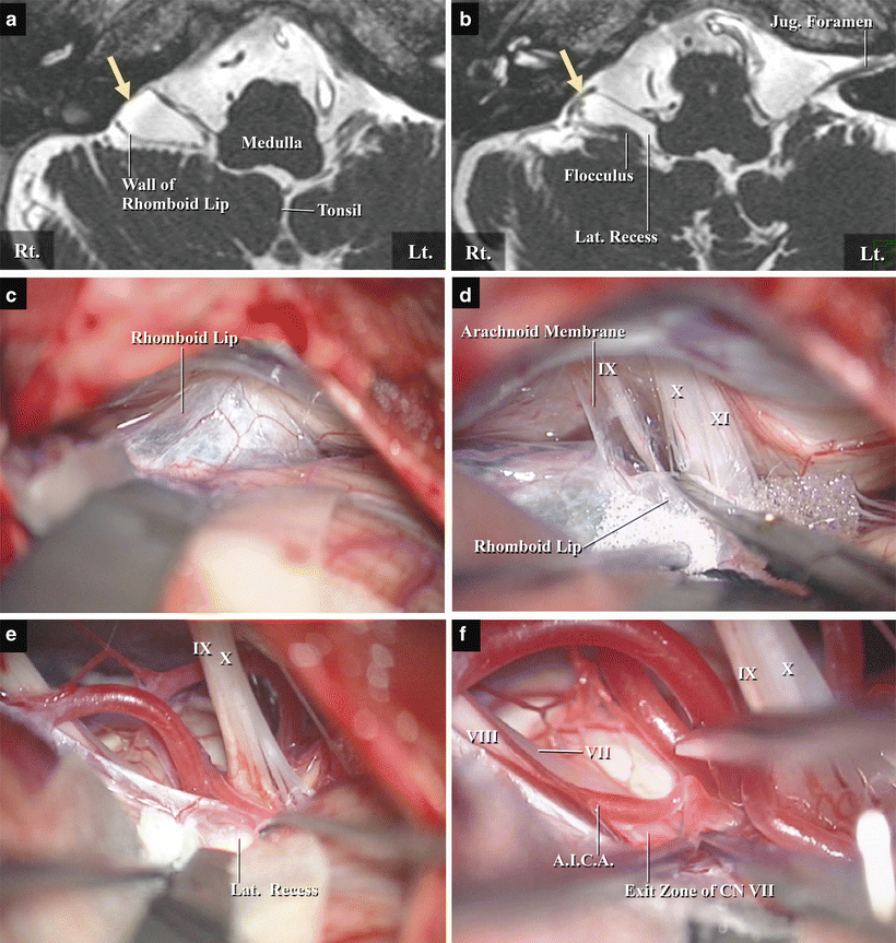
A 60-year-old female presented with right-sided HFS, which had persisted for 3 years. Neurological examination demonstrated typical HFS. Preoperative images revealed a common trunk-type anterior inferior cerebellar artery (AICA) and posterior inferior cerebellar artery (PICA) and that the loop of AICA was compressing REZ of CN VII. A cystic structure was observed ventral to the flocculus and in front of the jugular foramen, which continued medially to the lateral recess (Fig. 12.3a, b). The lateral recess of the affected side was dilated in comparison with the contralateral side (Fig. 12.3b). We therefore performed MVD surgery. When the cerebellomedullary cistern was exposed, an extremely large rhomboid lip was observed, which was adherent to the dorsal sides of CNs IX and X (Fig. 12.3c). The membrane of the rhomboid lip was tough and thick and contained small vessels. We separated the rhomboid lip membrane from the arachnoid membrane and the lower CNs. After separation, the arachnoid membrane was confirmed between the membrane of the rhomboid lip and the lower CNs (Fig. 12.3d). After dissection of the arachnoid membrane away from the lower CNs, the rhomboid lip could be retracted to the medial petrosal cerebellar surface. The choroid plexus protruding from the foramen of Luschka and the interior of the lateral recess were identified after incision of the rhomboid lip membrane (Fig. 12.3e). After these dissections, REZ of CN VII was clearly visible between the flocculus and the origin of CN IX (Fig. 12.3f). The offending artery was carefully detached from REZ, and small pieces of Teflon® felt were placed between the artery and the brainstem. HFS resolved after surgery, and the postoperative course was uneventful with no new deficits. This case was reported in Journal of Neurosurgery [21].


Fig. 12.3
The rhomboid lip on magnetic resonance imaging (MRI) scans and intraoperative photos of a representative case with a large rhomboid lip (from Nakahara Y et al. [21] with permission). (a) T2-weighted image of preoperative MRI revealing a cystic structure ventral to the flocculus and in front of the jugular foramen (yellow arrow). (b) MRI showing the cystic structure continuing medially to the dilated lateral recess. (c) A large rhomboid lip, wrapped around the jugular foramen and adherent to the lower CNs, was evident. (d) The rhomboid lip was dissected and moved to the medial petrosal (lateral) cerebellar surface. After separation of the rhomboid lip, the arachnoid membrane was observed. (e) The interior of the lateral recess and foramen of Luschka were exposed after incision of the rhomboid lip. (f) The offending artery, AICA, was exposed and detached from the REZ of CN VII
12.4 Surgical Procedures and Techniques for the Infrafloccular Approach
12.4.1 Positioning
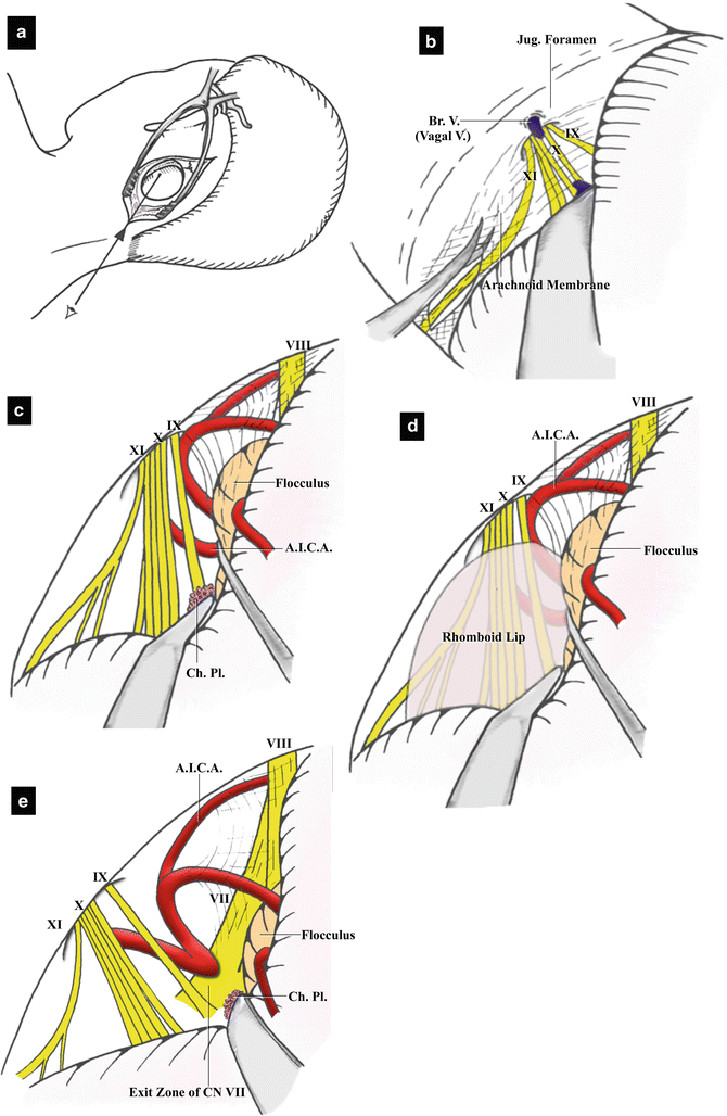
The patient is placed in a lateral park bench position and the head is fixed using three pins of a head fixation device to ensure that the head can be rotated during surgery. The pins should be placed at sufficient distance from the skin incision to avoid skin retraction. The head is rotated slightly to the contralateral side with anterior flexion to ensure that REZ of CN VII can be observed from the caudal side of the flocculus (Fig. 12.4a) [7, 10]. The patient’s shoulder on the lesion side should be caudally retracted, to avoid interference with the surgeon’s arm during surgery, within a limit that avoids brachial plexus palsy. The patient’s arm on the other side should be dropped from the operating table.


Fig. 12.4
Illustrations of the infrafloccular approach (from Matsushima T [7] with permission). (a) Skin incision and bony opening. The skin incision should be curved from the superolateral to the inferomedial side. The center of the bony opening should be at the posterior end of the mastoid incisura. The bony opening should be made below the mastoid incisura because the surgeon’s view will be from the inferoposterior side. (b) Opening of the cerebellomedullary cistern and lateral CMF. The cerebellomedullary cistern should be opened completely. When bridging veins are encountered around the jugular foramen, they should be coagulated before bleeding. The width of opening of the CMF depends on the case. (c) Dissection between CN IX and the flocculus and exposure of the REZ of CN VII. The tip of the spatula is placed on the choroid plexus originating from the foramen of Luschka. The flocculus and CN IX are completely dissected to create a space between them. (d) The rhomboid lip covers the lower CNs and prevents visualization of REZ of CN VII. In some cases, the rhomboid lip is large and adherent to the lower CNs. It should be separated from the lower CNs to create a space between CN IX and the flocculus. (e) Observation around REZ of CN VII. When the flocculus is retracted superiorly, the proximal portion and REZ of CN VII are easily observed. Offending arteries can then be identified
12.4.2 Skin Incision and Craniotomy
A curved linear skin incision is made along and just medial to the hairline, and the inferior half of the incision should be directed toward to the midline as a curved line to ensure that the skin flap does not interfere with the surgeon’s view through the surgical microscope [7, 14


Stay updated, free articles. Join our Telegram channel

Full access? Get Clinical Tree


