(1)
Department of Neurosurgery Faculty of Medicine, Saga University, Saga, Japan
Keywords
Glossopharyngeal neuralgiaOffending posterior inferior cerebellar artery and anterior inferior cerebellar arterySurgical approachesTranscondylar fossa approachSelection of an approach depending on arteries affected13.1 Introduction
Glossopharyngeal neuralgia (GPN) is one of several microvascular compression syndromes. The neuralgia is characterized by unilateral, paroxysmal, lancinating pain in the oropharynx or tonsillar fossa, spreading to the external auditory canal through the eustachian tube. It is often caused by activities such as talking, eating, swallowing, and coughing. “Typical” GPN is easily diagnosed when a patient presents the aforementioned symptoms in combination with radiological features, ascertained by magnetic resonance imaging (MRI)/magnetic resonance angiography (MRA), such as posterior inferior cerebellar artery (PICA) with a high origin that forms an upward loop (Fig. 13.1). However, unless these typical features of PICA are evident on MRI/MRA, the diagnosis and efficacy of microvascular decompression (MVD) remain uncertain. In such cases, the characteristics of the pain, e.g., location within the distribution of cranial nerve (CN) IX, paroxysmal, and association with swallowing, aid diagnosis. Carbamazepine therapy is effective and can be used for a diagnostic trial. Furthermore, the application of 10 % cocaine or lidocaine to the pharynx, a test block, relieves the pain of GPN. With the confirmation of these characteristics, the diagnosis of GPN becomes more reliable and successful outcomes of MVD can be expected.
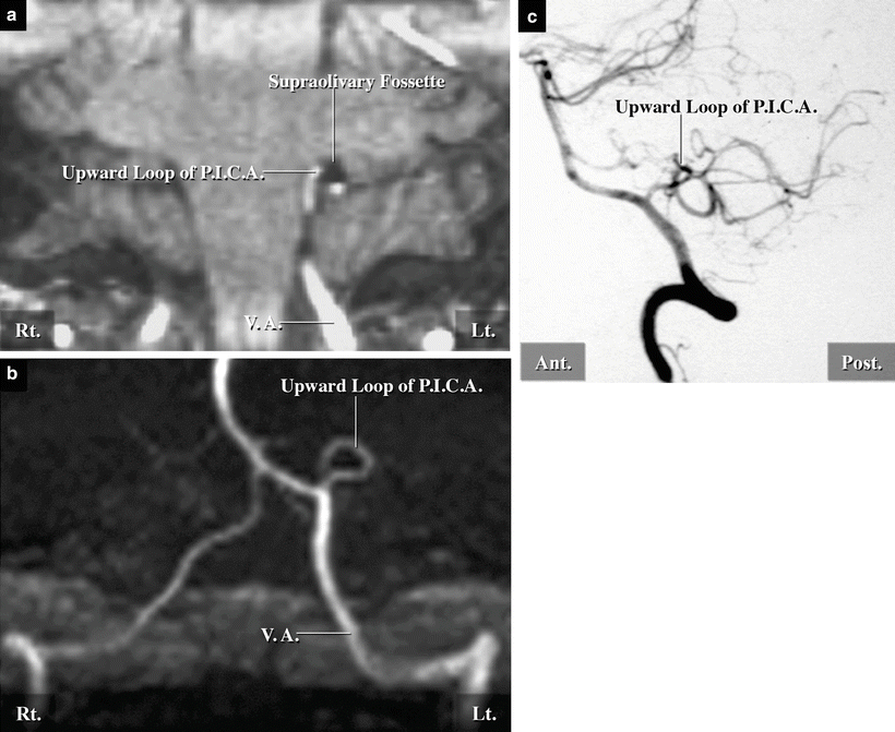
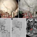
Fig. 13.1
Preoperative image analyses in a typical case of GPN with offending PICA. (a) Coronal section of an MRI at the level of the medulla oblongata showing left PICA coursing in the supraolivary fossette, forming an upward loop (red arrow) (from Matsushima T [10] with permission). (b) Anteroposterior view of an MRA image showing the anterior medullary segment of left PICA with a high origin forming an upward loop pointing superoposteriorly in the supraolivary fossette (red arrow). (c) Lateral view of the vertebral angiogram showing PICA forming an upward loop (red arrow) (from Matsushima T [10] with permission)
We have recently adopted two different approaches for MVD for GPN, depending on the offending artery [2]. In this chapter, we explain these approaches through the presentation of two typical cases, one with offending PICA and another with offending anterior inferior cerebellar artery (AICA). However, we focus on the case with offending PICA because it is the offending artery in most cases of GPN [4, 5, 11, 16, 17]. The development of diagnostic imaging techniques has allowed identification of the offending arteries and precise simulation of their surgical views prior to surgery. These techniques are helpful in selecting a suitable approach in each patient.
13.2 Offending Arteries of GPN (PICA or AICA) and Their Relationships to the Glossopharyngeal Nerve (CN IX)
PICA is the offending artery in over 80 % of cases of GPN caused by vascular compression and AICA or vertebral artery (VA) in the remainder [4, 5, 11, 16, 17]. We surgically treated 22 cases of GPN, including 18 with offending PICA (82 %) and four with offending AICA.
Normally, the lateral medullary segment of PICA seldom reaches the level of CN IX [6]. However, in GPN, PICA with a high origin forms an upward loop, coursing around the cisternal portion of CN IX near the supraolivary fossette, thereby compressing the nerve (Fig. 13.2) [3, 4, 10, 11]. These findings can be detected on preoperative MRI/MRA and represent the typical radiological features of GPN (Fig. 13.1).
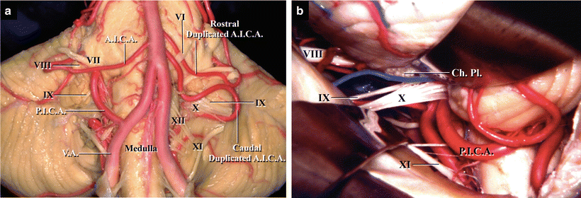
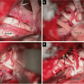
Fig. 13.2
Relationships between PICA, AICA, brainstem, and lower CNs in autopsied specimens. (a) The brainstem with PICA and AICA, anterior view. PICA on the right side originates from high on VA and forms an upward loop in the supraolivary fossette. On the left side, there are duplicated AICAs; the caudal one courses across the lower CNs in a rostral-to-caudal direction. (b) Surgical view of the left CMC, posterior view. Left PICA forms an upward loop in the supraolivary fossette and courses under the nerve roots of CNs IX and X
On the other hand, when the offending artery is AICA, a branch (usually, the caudal trunk) runs across CN IX in a cranial-to-caudal direction, compressing the nerve (Fig. 13.2a). Adhesion of the arachnoid membrane or choroid plexus to CN IX can also cause GPN.
13.3 Selection of Surgical Approach Depending on the Offending Artery
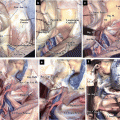
MVD for GPN is generally performed through the conventional lateral suboccipital approach (LSA), but the most suitable approach and positioning should be selected on the basis of the offending artery. In order to transpose an offending artery, it is necessary to pull the loop of offending PICA caudally, and offending AICA rostrally, from CN IX [2]. When the offending artery is PICA, its upward loop must be approached from the posteroinferior aspect through the transcondylar fossa (trans-CF) approach (Fig. 13.3) [4, 10, 11]. Furthermore, the approach is now combined with opening of the unilateral cerebellomedullary fissure (CMF), which is performed in the modified semiprone park bench position or the prone position [12]. The combined approach provides good visualization of CN IX and a wide operative field in the cerebellomedullary cistern (CMC) for transposing the offending PICA caudally [2, 12].
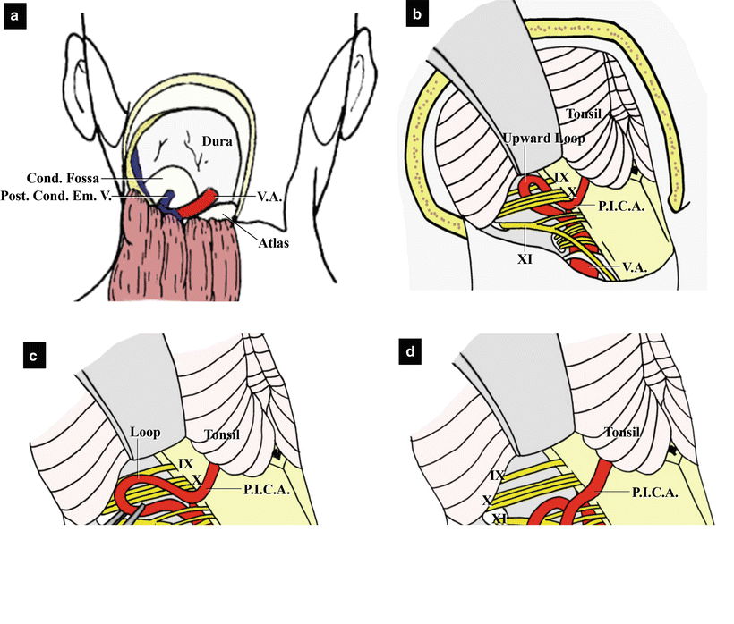
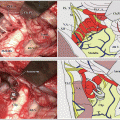
Fig. 13.3
Illustrations showing the transcondylar fossa approach and stitched sling retraction technique of MVD for GPN. (a) The skin flap together with the muscle has been detached from the suboccipital cerebellar surface. The posteroinferior portion of the occipital bone has been removed, leaving only the condylar fossa (from Matsushima T [9] with permission). (b) The entire courses of the cisternal portions of the lower CNs and offending PICA can be seen (from Matsushima T et al. [14] with permission). (c) The loop of PICA pulled out for transposition (from Matsushima T et al. [14] with permission). (d) PICA loop is then fixed to the dura mater using the stitched sling retraction technique (from Matsushima T et al. [14] with permission)
In cases of GPN caused by AICA, the offending artery must be transposed rostrally after opening the lateral CMF through the LSA, which is performed in the park bench position with the head turned to the lesion side or in the semiprone park bench position. The approach is therefore from a more inferior aspect than the conventional LSA. Opening of the lateral CMF facilitates the retraction of the biventral lobule and provides a wider operative field around the lower CNs in the CMF. The two approaches both provide adequate operative space to visualize the entire course of CN IX and transpose offending AICA or PICA. Selection of the best approach for each case allows the safe, easy, and complete transposition of an offending artery and reduces the risk of injury of the cerebellar hemisphere and/or lower CNs. Finally, it leads to a good outcome following MVD.
High-quality visualization via three-dimensional (3D) fusion imaging techniques has recently elucidated the 3D relationships between the offending artery and other important vessels, CNs, and brain structures. Their use preoperatively helps in the selection of a suitable approach for MVD [2].
13.4 Surgical Approach and Procedures for Offending PICA: The Trans-CF Approach Combined with Opening of the Unilateral CMF
We will now explain the approach and procedures of MVD for cases of GPN with offending PICA and present a representative case with intraoperative illustrations.
13.4.1 Craniotomy and Dural Opening
The bony opening should be large compared with that used during the LSA for hemifacial spasm (HFS). It should extend inferiorly to expose the inferolateral margin of the suboccipital (posterior) cerebellar surface. We usually utilize the trans-CF approach, in which the condylar fossa is removed (Fig. 13.3a) [4, 10, 11, 13, 14]. After the dura mater is opened, the inferior margin of the cerebellar hemisphere and foramen of Magendie should be exposed for dissection of the ipsilateral CMF (Fig. 13.3b).
Regarding the positioning, skin incision, craniotomy, and dural opening, refer to Chap. 7: “Microsurgical Anatomy of the Cerebellomedullary Fissure and Variations of the Transcerebellomedullary Fissure Approach” and Chap. 17: “Surgical Anatomy of and Approaches Through the Lateral Foramen Magnum: The Transcondylar Fossa and Transcondylar Approaches.”
13.4.2 Opening of the Lateral CMF and Observation of CN IX and the Surrounding Area
First, small bridging veins that obstruct the operative approach should be coagulated and cut before they are injured. Second, the ipsilateral CMF is dissected and opened to some extent, to make retraction of the inferolateral portion of the cerebellar hemisphere easy and safe (Fig. 13.3b) [12
 The Veins of the Posterior Cranial Fossa: Nomenclature
 The Cerebellopontine Angle: Basic Structures and the “Rules of Three”
 Microvascular Decompression Surgery for Hemifacial Spasm: The Lateral Suboccipital Infrafloccular Approach
 Occipital Artery–Posterior Inferior Cerebellar Artery Bypass Surgery
 Microsurgical Anatomy of and Surgical Approaches to the Jugular Foramen
 Microsurgical Anatomy of the Cerebellomedullary Fissure and Variations of the Transcerebellomedullary Fissure Approach


Related posts:
The Veins of the Posterior Cranial Fossa: Nomenclature
The Cerebellopontine Angle: Basic Structures and the “Rules of Three”
Microvascular Decompression Surgery for Hemifacial Spasm: The Lateral Suboccipital Infrafloccular Approach
Occipital Artery–Posterior Inferior Cerebellar Artery Bypass Surgery
Microsurgical Anatomy of and Surgical Approaches to the Jugular Foramen
Microsurgical Anatomy of the Cerebellomedullary Fissure and Variations of the Transcerebellomedullary Fissure Approach
Stay updated, free articles. Join our Telegram channel
Full access? Get Clinical Tree
