(1)
Department of Neurosurgery Faculty of Medicine, Saga University, Saga, Japan
Keywords
Cerebellomedullary fissureTranscerebellomedullary fissure approachTelovelar approachSubtonsillar approachFourth ventricular surgeryCerebellomedullary cistern7.1 Introduction
Figure 7.1 is an illustration of an the Cerebellomedullary Fissure and Variations…?>intraoperative view of a distal posterior inferior cerebellar artery (PICA) aneurysm. Some neurosurgeons must have had their own experiences of opening the cerebellomedullary fissure (CMF) during surgery, such as the clipping of the distal PICA aneurysm, before our first report of the transcerebellomedullary fissure approach (trans-CMF approach) [10, 21]. The CMF is the biggest cleavage plane of the three cerebellar–brainstem fissures. Opening the CMF is useful during various surgeries in the lower posterior cranial fossa in the same way as opening the Sylvian fissure in supratentorial surgeries. We proposed the trans-CMF approach as an approach utilizing the opening of any part of the CMF and have developed several variations [9, 10, 12, 13, 20–22, 26, 30, 31, 33, 34, 36]. In this chapter, the anatomy of the CMF, the evolution and concept of the trans-CMF approach, and the variations of the approach, such as the medial and lateral routes and the unilateral trans-CMF approach, are explained.
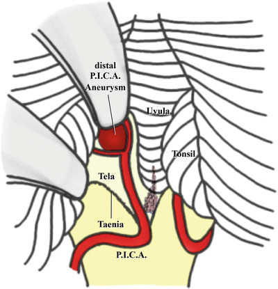
Fig. 7.1
Illustration of an intraoperative view of surgery for a left distal posterior inferior cerebellar artery aneurysm through the trans-CMF approach (from Matsushima T [18] with permission)
7.2 Evolution of the Transcerebellomedullary Fissure Approach with the Opening of the CMF
While studying the fourth ventricle in cadaveric specimens in the early 1980s, Rhoton AL Jr and I observed a large fissure between the cerebellum and medulla, which we named the CMF [37]. After I started performing surgeries, I encountered an unforgettable case of a fourth ventricular tumor, for which I used the transvermian approach. I thought I could remove the tumor completely, but it recurred in the left lateral recess 1 year after surgery [19, 21]. I recognized that visualization of the lateral recess through the transvermian approach was insufficient and thenceforth started to dissect the CMF almost entirely to expose the entire fourth ventricle, particularly the lateral recess. In the early 1990s, based on these surgical experiences, we reported the usefulness of opening the CMF instead of splitting the inferior vermis during fourth ventricular tumor surgeries [21, 22].
Since our proposal, it has become established that opening the CMF during fourth ventricular surgery yields a wide operative field and decreases the incidence of vermal split syndrome (cerebellar mutism syndrome) and residual tumors in the lateral recess. In the late 1990s and early 2000s, the medial route of the approach was further developed by several authors [4, 10, 11, 14, 21, 26, 30, 33, 34, 41, 43, 47, 48]. The effects of opening the CMF through the medial route, also called the telovelar approach, were also evaluated by cadaveric studies [3, 39, 44].
The approaches and procedures for opening the CMF have been reported under several names because they were applied to different lesions in different areas of the fourth ventricle and different portions of the CMF were opened and utilized. They include the median inferior suboccipital transfissural [46, 47], CMF [14], subtonsillar–trans-CMF [48], telovelar [39], and subtonsillar [11, 45] approaches. The subtonsillar approach corresponds to the lateral recess-type medial route or unilateral trans-CMF approach as per our classification [30, 31].
Because opening the CMF provides wide operative fields both inside and around the fourth ventricle, we have extended its applications to many surgeries for various lesions [9, 12, 13, 18, 30, 31, 36]. First, we developed the medial route for tumors mainly occupying the fourth ventricle surgically removed via the midline suboccipital approach; we subsequently developed the lateral route, in combination with the transcondylar fossa approach from the lateral foramen magnum side [12, 36]. We have recently used the trans-CMF approach during surgeries for lesions, such as vascular lesions, in the cerebellomedullary cistern (CMC) [13, 31]. This approach can also be used for tumor surgeries.
7.3 Microsurgical Anatomy of the CMF
7.3.1 Neural Structures
The CMF is a cleavage plane between the cerebellar hemisphere and medulla oblongata and is usually closed by arachnoidal adhesion (Fig. 7.2a) [23, 24, 27, 35, 37, 42]. The roof of the CMF is formed by the tonsil and biventral lobule, and the floor by the posterior surface of the medulla oblongata and tela choroidea, which is the caudal half of the inferior part of the fourth ventricular roof (Fig. 7.2b–e). The CMF comprises three main spaces (Fig. 7.2a, b):
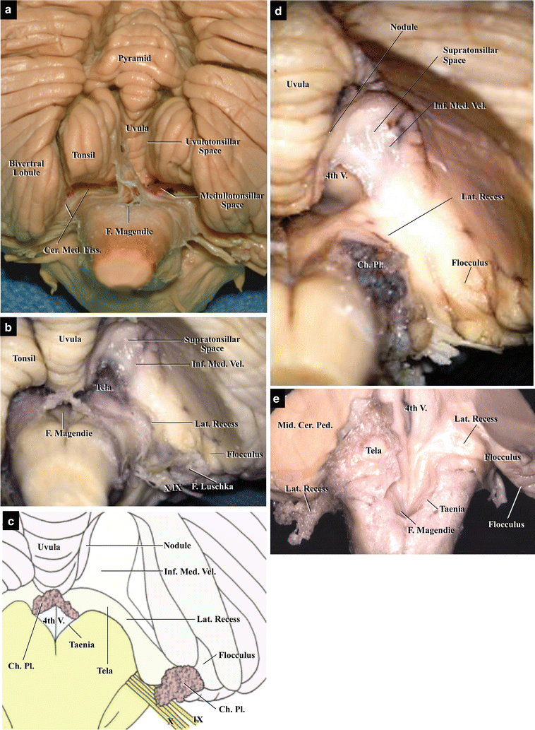
Fig. 7.2
Anatomy of the neural structures of the CMF: step-by-step dissection. (a) Suboccipital cerebellar surface, posterior view. The uvulotonsillar and medullotonsillar spaces can be seen (from Matsushima T [18] with permission). (b) Exposure of the right half of the CMF, posterior view. The right tonsil has been removed, exposing the floor of the CMF, which is also the roof of the fourth ventricle, on the right side. The supratonsillar space is also visible (from Matsushima T [18] with permission). (c) Illustration of Fig. 7.2b showing the floor of the right CMF after removal of the tonsil. The inferior medullary velum, tela choroidea, and lateral recess are visible. (d) Exposure of the interior of the fourth ventricle. The tela choroidea has been reflected on the right side, revealing the interior of the ventricle (from Matsushima [18] with permission). (e) Relationship between the CMF and the floor of the fourth ventricle. The cerebellar hemispheres and right half of the fourth ventricular roof, which is the inferior medullary velum and tela choroidea, have been removed
The CMF extends from the uvulotonsillar space near the midline to the CMC laterally (Fig. 7.3a, b). The CMC is a cistern surrounded by the jugular tubercle laterally and medulla oblongata medially. The lateral recess, a part of the fourth ventricle, extends into the upper CMC and is situated on the roots of cranial nerves (CNs) IX and X. The CMF is continuous with the CMC, and their transition is gradual.
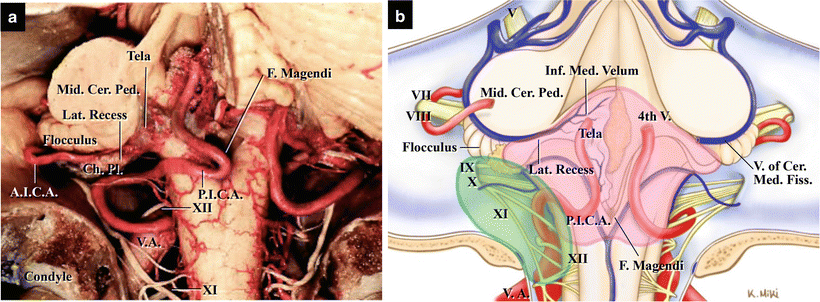
Fig. 7.3
Relationships between the CMF and CMC (from Matsushima T et al. [31] with permission). (a) Cadaveric specimen, posterosuperior view. The left cerebellar hemisphere has been removed, revealing the fissure and cistern. (b) Illustration of the relationships. The red encircled area is the CMF and the blue encircled area is the CMC. They gradually merge
In the midline, the uvula and nodulus are sandwiched between the tonsils (Fig. 7.2a). The tonsil is attached only at its superolateral portion, the tonsillar peduncle. Retracting the tonsil and biventral lobule superolaterally after detaching them from the surrounding structures exposes the floor of the CMF, which is the fourth ventricular roof (Fig. 7.2b, c). The inferior medullary velum, which forms the cranial half of the inferior part of the fourth ventricular roof, is a membranous, translucent layer connecting the nodulus and flocculus (Fig. 7.2d). The tela choroidea with the choroid plexus is a membrane attached to the medulla oblongata inferolaterally by the taenia (Fig. 7.2c–e). When the tela choroidea is resected and reflected, the floor of the fourth ventricle is revealed (Fig. 7.2d). The lateral recess is narrow, comprising curved pouches located at the lateral angles of the floor of the fourth ventricle (Fig. 7.2c–e). The lateral recess is formed by the rhomboid lip and tela choroidea, which extend laterally from the wide middle portion of the fourth ventricle into the CMC. The choroid plexus is suspended from the ventricular surface of the tela choroidea and protrudes into the cerebellopontine angle through the foramen of Luschka (Fig. 7.2c–e). The lower CNs run below the lateral recess. In some cases, a large rhomboid lip is adherent to the lower CNs [5, 40]. The middle cerebellar peduncle and flocculus are situated just anterosuperior to the lateral recess and form the rostral limit of the lateral part of the CMF (Fig. 7.2e).
7.3.2 Vascular Structures
The main artery running in the CMF is PICA, but the anterior inferior cerebellar artery (AICA) also passes near the lateral recess (Fig. 7.4a, b) [18, 25, 37, 42]. PICA, arising from the vertebral artery (VA), runs around the medulla oblongata, forms a caudal loop near the foramen of Magendie, and runs in the CMF as the tonsillomedullary segment. The tonsillomedullary segment of PICA gives rise to perforating arteries to the medulla oblongata. The medial part of the tela choroidea is supplied by the choroidal arteries from PICA and the lateral part is supplied by those arising from AICA. PICA forms a cranial loop in the CMF by coursing around the superomedial surface of the tonsil, exits the CMF, and bifurcates into vermian (medial) and tonsillohemispheric (lateral) branches.
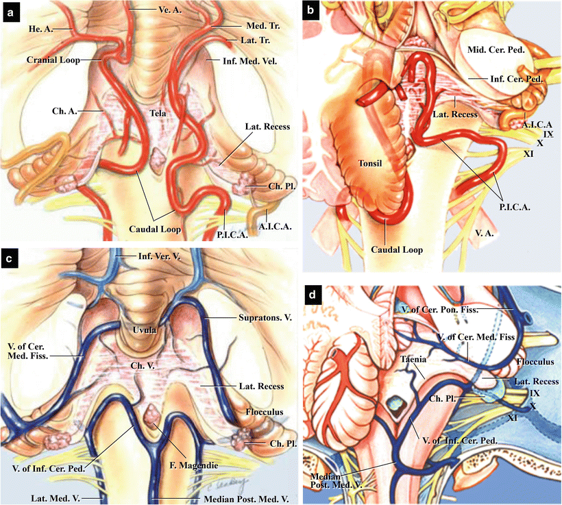
Fig. 7.4
Color illustrations showing arteries and veins in the CMF. (a) Arteries in the CMF, posterior view. PICAs course in the CMF and form caudal and cranial loops (modified from Matsushima T et al. [37] with permission). (b) Arteries in the CMF, superior view. The course of PICA in the CMF and its relationship with the fourth ventricle and tonsil (modified from Matsushima T et al. [37] with permission). (c) Veins in the CMF, posterior view. The main draining veins in the CMF are the bilateral veins of the CMF, which course on the inferior medullary velum and flocculus (modified from Matsushima T et al. [37] with permission). (d) Veins in the CMF, superior view. The courses of the veins of the CMF and of the inferior cerebellar peduncle in the CMF (modified from Matsushima T et al. [38] with permission)
The main veins running in the CMF are the vein of the cerebellomedullary fissure and the vein of the inferior cerebellar peduncle (Fig. 7.4c, d) [23, 25, 29, 37, 38]. The vein of the cerebellomedullary fissure receives venous drainage from the lateral wall and roof of the fourth ventricle and courses laterally on the lateral recess to drain into the petrosal draining group of the veins of the posterior cranial fossa. The vein of the inferior cerebellar peduncle runs on the inferior cerebellar peduncle along the inferolateral margin of the fourth ventricle. It connects with the petrosal draining group superolaterally and the median posterior medullary vein posteromedially. The vein of the inferior cerebellar peduncle is a useful anatomical landmark during incision of the taenia, which is the inferolateral margin of the fourth ventricular roof.
7.4 Classification of the Trans-CMF Approach (the Medial and Lateral Routes and the Unilateral Trans-CMF Approach)
The CMF is hidden from posterior view by the cerebellar hemisphere. However, this cleavage is continuous to the CMC laterally and the cisterna magna inferiorly (Fig. 7.5a, b). Fourth ventricular ependymomas or medulloblastomas often extend inferiorly and laterally through the CMF (Fig. 7.5c, d). As indicated by two arrows in Fig. 7.5b, fourth ventricular tumors usually grow and extend inferiorly and laterally into the spinal canal and CMC through the CMF. Therefore, surgeons can approach the interior of the fourth ventricle both from the midline and lateral side (Fig. 7.5e). This is the basic concept behind the medial and lateral routes of the trans-CMF approach [12, 18, 31, 36]. In the medial route, the tonsil can be elevated superiorly or superolaterally, although only lateral retraction is possible in the transvermian approach. In the lateral route, the biventral lobule and/or tonsil are mainly retracted superiorly after opening the lateral CMF. After surgical experience of using both routes in the early 2000s, we started to intentionally open the unilateral normal CMF to obtain a wide operative field in the CMC, especially during vascular surgeries for the treatment of VA–PICA aneurysms and during microvascular decompression for glossopharyngeal neuralgia (GPN) [9, 13, 31]. We designated this approach, which combines the medial and lateral routes, the unilateral trans-CMF approach [31]. We explain the trans-CMF approach by separating it into these three approaches because we believe this classification helps in understanding the approach and its variations; however, there are many variations of the three approaches. For example, similar to opening the Sylvian fissure, opening some parts of the CMF is very useful during many different surgeries in the lower posterior cranial fossa, from the fourth ventricle to the CMC.
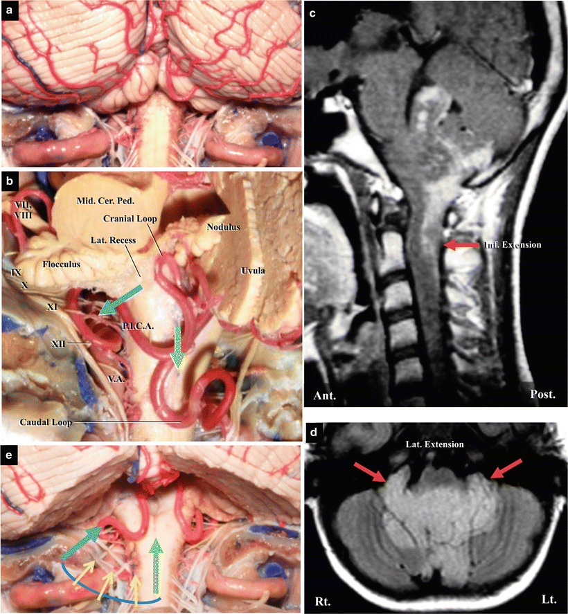
Fig. 7.5
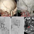
Extensions of fourth ventricular tumors and variations of the approaches to the fourth ventricle (modified from Matsushima T [18] with permission). (a) Autopsy specimen demonstrating the suboccipital cerebellar surface and medulla oblongata, posterior view. The CMF is hidden by the bilateral cerebellar hemispheres. (b) Autopsy specimen demonstrating the extension of the tumors, left posterolateral view. The left cerebellar hemisphere has been removed, exposing the left CMF. The green and blue arrows indicate inferior and inferolateral extensions of fourth ventricular tumors through the CMF. (c) MRI of a case of ependymoma, sagittal view. The ependymoma extends inferiorly to the level of C2 through the CMF. (d) MRI of the same ependymoma, axial view. The ependymoma extends laterally to the CMCs through the CMF. (e) Autopsy specimen demonstrating approaches to the fourth ventricle, posterior view. The tonsils and parts of the biventral lobules have been removed, exposing the bilateral CMFs. The green and blue arrows indicate the medial and lateral routes, respectively. The fourth ventricle can be approached not only from the midline (green arrow) but also from the lateral side (blue one). Yellow small arrows also indicate the approaches from the posterolateral sides to the fourth ventricle
7.5 Four Regions Exposed Through the Trans-CMF Approach
When we open the CMF, it is possible to expose four different regions in or around the fourth ventricle (Fig. 7.6):
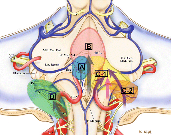
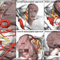
Fig. 7.6
Illustration of four regions exposed by the trans-CMF approach (from Matsushima T et al. [31] with permission). A (sky blue), intra-CMF lesions; B (blue), intra-fourth ventricular lesions; C (C-1, C-2), lesions from the CMF to the CMC; C-1 (yellow), CMF lesions with lateral extensions; C-2 (orange), CMC lesions with medial extensions; and D (green), intra-CMC lesions. The purple arrows show the lateral recess type of the medial route and the lateral route. The pink arrow indicates the unilateral trans-CMF approach, which is the combination of the two routes
Region A: the interior of the CMF/outside of the ventricle (for intra-CMF lesions)
Region B: the interior of the fourth ventricle, particularly the medial portion (for intra-fourth ventricular lesions)
Region C: the region from the CMF to the CMC (for CMF and CMC lesions)
Region D: only the CMC (for intra-CMC lesions) [31]
Region A (intra-CMF) is used for distal PICA aneurysms or tumors such as hemangioblastoma [6, 21]. The occipital artery (OA)–PICA anastomosis was recently reported in this region after opening the CMF [1]. Region B (intraventricular) targets tumors such as ependymoma occupying the ventricle or cavernoma in the ventricular floor. Region C (CMF and CMC) targets ependymoma, medulloblastoma, choroid plexus papilloma, epidermoid tumor, lower CN neurinoma, and jugular tubercle meningioma. Region C is subdivided into C-1, for a main lesion in the CMF (CMF lesions with lateral extension), and C-2, for a main lesion in the CMC (CMC lesions with medial extension). In Region D (intra-CMC), we have observed VA–PICA aneurysm and GPN.
7.6 Medial Route of the Trans-CMF Approach: Procedures and Maneuvers
In the medial route, the midline suboccipital approach is used and an operative microscope is inserted from the medial side. The medial portions of the CMF and fourth ventricle are exposed by retracting the tonsils superolaterally after dissecting the CMF. The medial route is applied for intra-CMF (Region A) lesions, intraventricular (Region B) lesions, and lesions in the CMF with lateral extension into the CMC (Region C1). First, we will explain the benefits of and exposure gained through the use of the medial route; then we will describe its surgical procedures and maneuvers in detail, while presenting three representative cases operated using this route.
7.6.1 Advantages of and Exposures Gained Through Use of the Medial Route
The medial route has the following advantages. First, opening the CMF makes retraction of the cerebellar hemisphere (tonsil and/or biventral lobule) safe and pronounced and facilitates good visualization of the lateral portion of the fourth ventricle, including the lateral recess. Therefore, it reduces the risk of leaving a lateral portion of a tumor [21, 26, 30, 33, 34]. Second, the cerebellar mutism syndrome, a common complication caused by splitting the inferior vermis, is avoided in this approach [4, 7, 14, 30, 34]. Third, this approach enables coagulation of the feeding arteries of a tumor at the early stages of surgery, thereby reducing bleeding from the tumor [21, 30, 33, 34].
Regarding the exposures gained through use of the medial route, Tanriover et al., who conducted a study comparing the transvermian and telovelar approaches to the fourth ventricle in cadaveric specimens, stated that the transvermian approach provided slightly better visualization of the medial part of the superior half of the fourth ventricular roof, but the telovelar approach provided additional exposure of the lateral recesses and foramen of Luschka [44]. Our clinical experiences were almost identical [21, 30]: we were able to remove a cavernoma in the fourth ventricular floor near the cerebral aqueduct and an ependymoma in the left lateral recess [10, 19, 21], but unable to remove a choroid plexus papilloma near the fastigium. We did not need to incise the inferior medullary velum in any case except one of choroid plexus papilloma originating from the fastigium. In cases where a tumor is located around the fastigium or originates from the vermis, it may be necessary to switch from the trans-CMF approach to the transvermian approach.
7.6.2 Surgical Procedures
7.6.2.1 Positioning and Bony Opening
To begin with, the patient is placed in the Concorde position [15]. As the line of sight through the operative microscope must look into the fourth ventricle from the posteroinferior side after suboccipital midline craniotomy, the patient’s head is tilted anteriorly as much as possible [28, 33]. Regarding the bony opening, as already explained in Chap. 6: “Midline Suboccipital Approach and Its Variations for Fourth Ventricular or Cerebellar Hemispheric Lesions,” the size and shape of a bony opening depend on the location of a lesion. For example, for a lesion with lateral extension into the lateral recess or CMC, the lateral foramen magnum on the lesion side should be removed. If bone removal at the lateral foramen magnum is insufficient, the bony margin will become an obstacle to retract the tonsil. Therefore, the inferior margin of the tonsil should be exposed, and if necessary, laminectomy of C1 should be performed.
Related posts:
 The Veins of the Posterior Cranial Fossa: Nomenclature
 The Cerebellopontine Angle: Basic Structures and the “Rules of Three”
 Microvascular Decompression Surgery for Hemifacial Spasm: The Lateral Suboccipital Infrafloccular Approach
 Occipital Artery–Posterior Inferior Cerebellar Artery Bypass Surgery
 Microvascular Decompression for Glossopharyngeal Neuralgia: Surgical Approaches Depending on the Offending Artery
 The Retrosigmoid Lateral Suboccipital Approach: Basic Approach and Variations
Stay updated, free articles. Join our Telegram channel

Full access? Get Clinical Tree


