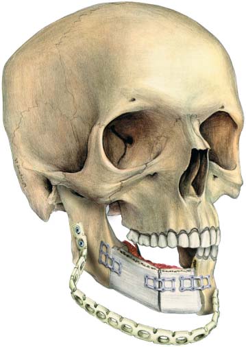
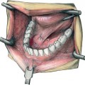
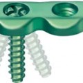
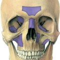
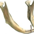
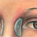
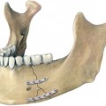
33 Mesh Fixation of Bone Different osteosynthesis techniques can be applied to fix bone grafts when reconstructing the mandible. This chapter looks at titanium mesh, which adds to the osteosynthesis techniques described in previous chapters and completes the spectrum of available fixation methods. Lambotte published an article in 1913, describing the treatment of a median mandibular fracture using a plate, which identifies the basic principles for mesh osteosynthesis. Boyne (1969) first used titanium mesh for mandibular reconstruction; later Hauenstein and Steinhäuser (1977) reported on technical advantages and clinical experiences. The titanium mesh was originally developed for mandibular reconstruction in combination with cancellous bone chips (Boyne, 1969). The idea was to replace the mandible by a U-shaped titanium mesh filled with autologous iliac crest spongiosa (Steinhäuser, 1982; Dumbach and Steinhäuser, 1983; Dumbach, 1985; Dum-bach et al., 1994). A range of titanium meshes has been developed, based on those used by earlier workers, and are available in a selection of different sizes. Wolff et al. (1996) reported on 24 cases of mandibular reconstruction using a fibula free flap with microsurgically anastomosed vascular pedicles. He observed one broken plate and one infection at the osteosynthesis line. Individual patient requirements determine which size of titanium mesh to use. For mandibular reconstruction, the 2 mm system is the osteosynthesis material of choice. In special cases where free bone of the alveolar crest, or other bone graft, is to be fixed on the mandible to reconstruct the alveolar ridge, the 1.5 mm system is useful (Figs. 33.1, 33.2). Further development of mesh fixation concerning the principles of three-dimensional fixation of the bone using titanium mesh has been introduced by Farmand (1991, 1995). He developed three-dimensional osteosynthesis plates for multipurpose use. Mesh systems are valuable in that they give the surgeon the opportunity to construct a plate according to the individual needs of the patient. With a special instrument, the mesh cutter, custom plates can be produced to meet the specific demands presented by the particular part of the facial skeleton to be reconstructed. Custom-made osteosynthesis plates can satisfy the surgeon’s individual needs for different approaches. Excellent results have been achieved using titanium mesh systems in the reconstruction defects of the facial skeleton during tumor surgery. Titanium mesh is ideal for use in the lateral orbit, or other bony structures of the facial skeleton, in interdisciplinary treatments for craniofacial tumors involving the neurosurgeon and the ophthalmologist (Fig. 33.3). This mesh reconstruction of the skeletal parts gives the patients an acceptable aesthetic and functional outcome. In interdisciplinary treatment of cranio-facial tumors, a mesh system is nearly always used as the osteosynthesis material for reconstruction of the resected bone. Fig. 33.1 Mandible reconstructed by microsurgically anastomosed iliac crest flap using 2 mm titanium mesh. Note the temporary fixation of the ascending rami during the osteosynthesis of the iliac crest flap.
Introduction
Related posts:
Stay updated, free articles. Join our Telegram channel

Full access? Get Clinical Tree


