Chapter 6 Table 6-1 Characteristics of nevus versus melanoma
Melanocytic neoplasms
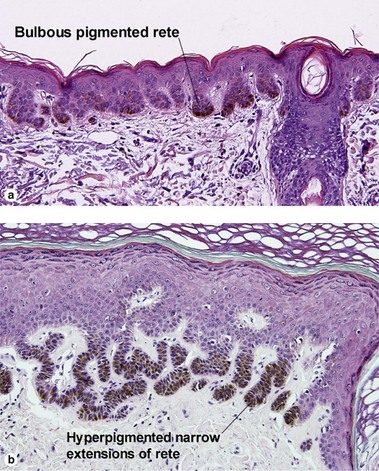
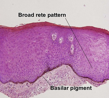
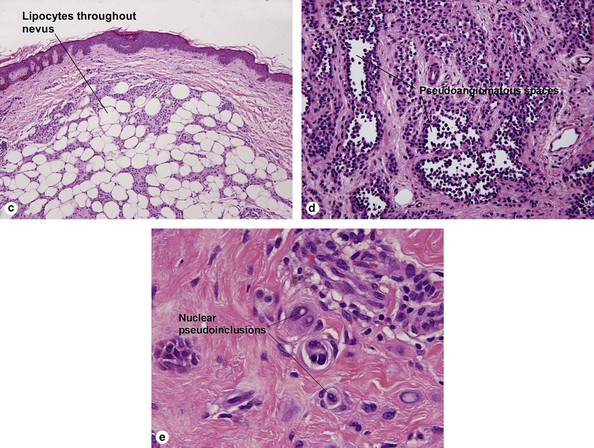
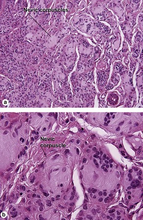
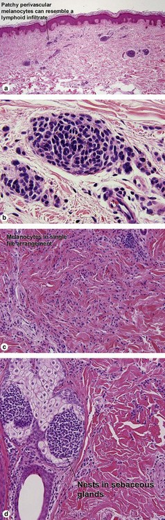
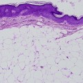
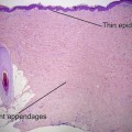
Benign melanocytic nevus
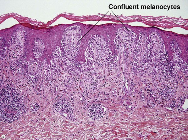
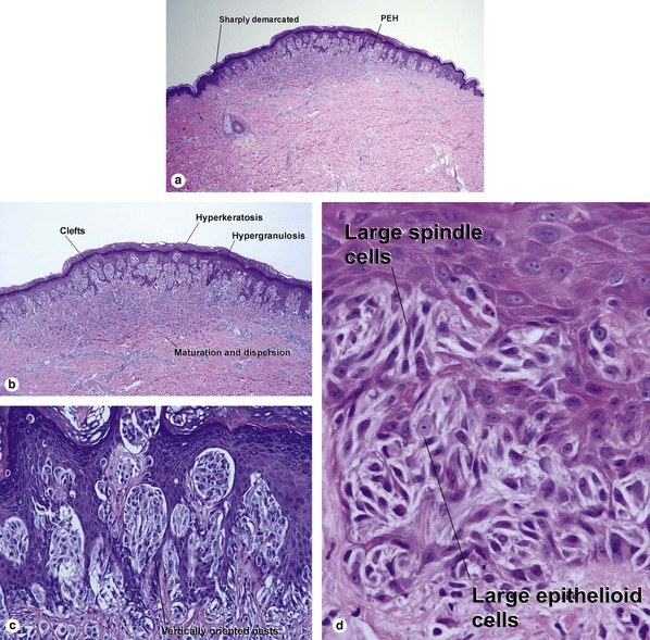
Table 6.1 gives general rules, and is a good starting point for the evaluation of pigmented lesions. There are exceptions to the rules. For example, blue nevi show no evidence of maturation or dispersion. They are commonly deeply pigmented to the base of the lesion. They are readily recognized by their wedge-like or bulbous outline and characteristic cytologic features.
Characteristic
Nevus
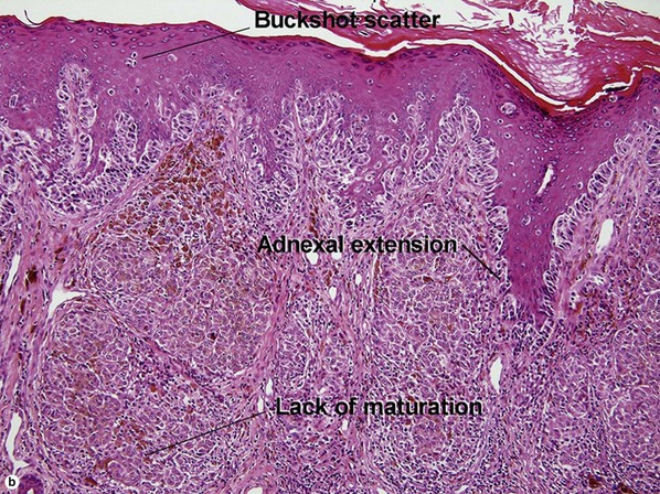
Melanoma
Lateral circumscription
Sharp
Variable
Bilateral (right to left) symmetry
Yes
Commonly asymmetrical
Top to bottom symmetry
No
Variable
Size
Small
Usually quite broad
Dermal–epidermal junction
Well nested
Non-nested melanocytes usually outnumber nests in areas
Shape of junctional nests
Round to oval
Often elongated and bizarre
Location of junctional nests
Tips and sides of rete
Tops of dermal papillae often involved as well
Spacing of junctional nests
Regular
Usually irregular
Buckshot scatter in epidermis
Absent except in the center of Spitz nevi, pigmented spindle cell nevi, acral nevi, traumatized nevi, and sunburned nevi
Variable (present in superficial spreading malignant melanoma, usually not prominent in lentigo maligna and acral lentiginous malignant melanoma)
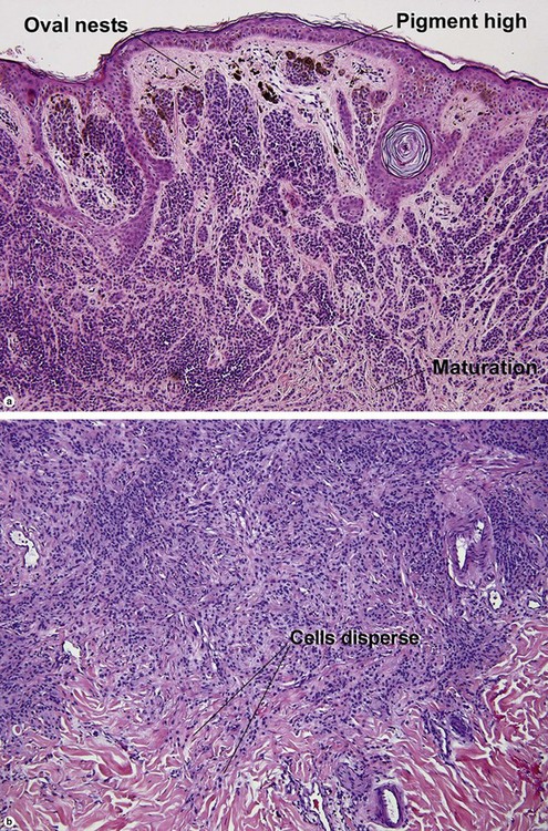
Maturation
Cells become smaller and more neuroid from top to bottom
Typically fails to mature
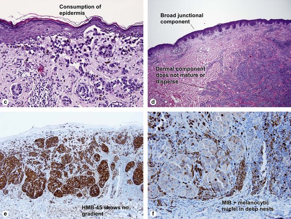
Dispersion
Disperses to single units at base of lesion
Generally remains nested at base
Junctional vs dermal nests
Dermal nests smaller than junctional nests; from top to bottom, nests become smaller, melanocytes disperse
Dermal nests often larger than junctional nests
Deep mitoses
Rare
Variable
Deep pigment
No
Variable
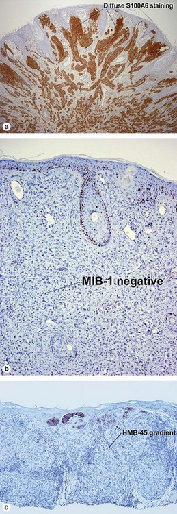
HMB-45
Top-heavy
Commonly stains strongly to base
MIB-1
No deep nuclei positive
Deep nuclei commonly positive
S100A6
Spitz nevi usually stain diffusely
Usually patchy