CHAPTER 7 Mechanism of Action of Sclerotherapy
General Mechanism for Producing Endothelial Damage
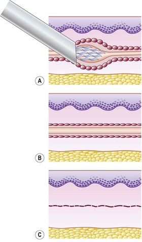
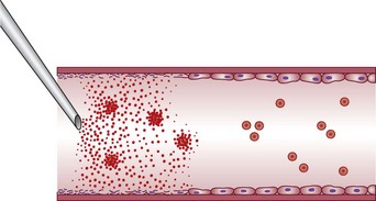
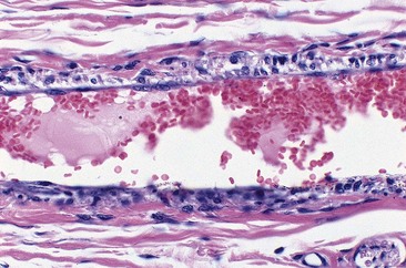
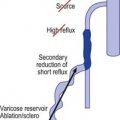
Sclerotherapy refers to the introduction of a foreign substance into the lumen of a vessel, aiming to create venous wall damage leading to occlusion of the vessel (Fig. 7.1). This procedure, when performed on telangiectasias, is referred to as microsclerotherapy.1
In addition, endothelial cells in one organ and in one location may act differently than endothelial cells elsewhere. They appear to be specialized to their area. Thus it is paradoxical that sclerotherapy treatment is not without significant adverse sequelae (see Chapter 8).
Endothelial destruction by sclerosing solutions is both dose and time dependent.6 In vitro studies of cultured endothelial cells demonstrated activation of calcium signaling and nitric oxide pathways followed by cell death after exposure to sclerosing solutions. Cell death occurred within 15 minutes with 0.3% polidocanol (POL) or 0.1% sodium tetradecyl sulfate (STS). At less than 0.003% POL or 0.005% STS cells remained alive after 60 minutes. Combined with protein-binding of detergent sclerosing solutions and a 10,000-fold dilution after release into the circulation, these studies demonstrate the safety of sclerotherapy, since sclerosing solutions are rapidly diluted to ‘safe’ concentrations distal to the point of injection.
Total endothelial destruction results in the exposure of subendothelial collagen fibers, causing platelet aggregation, adherence, and release of platelet-related factors. This series of events initiates the intrinsic pathway of blood coagulation by activating factor XII. Ideally, sclerosing solutions otherwise should not cause activation or release of thromboplastic activity because this would initiate the extrinsic pathway of blood coagulation. Excessive thrombosis is detrimental to the production of endofibrosis because it may lead to recanalization of the vessel as well as excessive intravascular and perivascular inflammation and its resulting sequelae (see Chapter 8). This is thought to be prevented or at least minimized with postsclerotherapy compression (see Chapter 6). However, thrombosis usually occurs to some degree as a result of sclerotherapy.
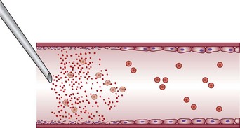
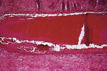
If a thrombus is formed, it should be well anchored to the venous wall to prevent embolization. Wolf7 in 1920 established that effective sclerosis causes thrombosis that penetrates the full thickness of the adventitia of the vessel wall. Schneider8 has shown in histologic examinations of sclerosed varices that the strongest fixation of a thrombus occurs in areas where the entire endothelium is destroyed. Therefore, endothelial damage must be complete and should result in minimal thrombus formation with subsequent organization and fibrosis (Fig. 7.2). In addition, after sclerotherapy, maximum full-thickness fibrosis of the treated segment occurs after 6 weeks of compression.9 Therefore, in addition to limiting the extent of thrombosis, compression may facilitate endofibrosis (see Chapter 6). Chleir and Vin10 have described the differences between a clot observed during a spontaneous thrombosis ‘thrombus’ and during the sclerosing process, for which they suggested the neologism ‘sclerus’ (Table 7.1).
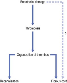
Table 7.1 Thrombus versus sclerosis: comparison of vein content during a thrombosis and during a sclerosing process
| Thrombus | Sclerosis | |
|---|---|---|
| Pathophysiology | Hematological phenomenon | Tissue phenomenon |
| Activation of Virchow’s triad | Parietal mechanism | |
| Recanalization without fibrosis | Fibrosis | |
| Clinical | Painful | More discomfort than pain |
| Inflammatory | No inflammation | |
| Indurate plaques | No indurate plaque | |
| Biology | Positive d-dimers | Negative d-dimers |
| B-mode ultrasound | Convex toward junction | Concave toward junction |
| ‘Rosette’ picture | Parietal thickening | |
| No parietal adhesion | Parietal adhesion | |
| Dilatation | Retraction | |
| Pathology | No parietal lesion | Inflammatory infiltration of venous wall |
| Thrombus rich in RBCs and platelets | Sclerosis contains more WBCs and less RBCs than thrombus | |
| Rare WBCs |
RBCs, red blood cells; WBCs, white blood cells.
From Chleir F, Vin F: Actualités Vasculaires Internationales 35:18, 1995.
Endothelial damage can be provoked by a number of mechanisms, such as a change in the surface tension of the plasma membrane or modification of the physical–chemical milieu of the endothelial cell through a change of intravascular pH or osmolality. The endothelium can be destroyed directly by caustic chemicals or by other physical factors such as heat and cold. For sclerotherapy to be effective without recanalization of the thrombotic vessel, the endothelial damage and resulting vascular necrosis must be extensive enough to destroy the entire blood vessel wall.11
Destruction of the entire vessel wall and not just the endothelium is necessary, as demonstrated in animal studies described later in this chapter. The reason may relate to the multifunctional nature of vascular smooth muscle cells. These cells, which are found in significant concentration within superficial veins (see Chapter 2), have a large number of functions, including the synthesis of collagen, elastin, and proteoglycans.12 It is hypothesized that if they remain viable, they can regenerate a foundation that promotes migration of undamaged adjacent endothelial cells that allow recanalization of the treated vessel.13,14
In addition, for effective destruction of a varicosity or telangiectasia, the entire vessel must be sclerosed to prevent recanalization. Recanalization occurs easily in vessels where only a section of endothelium is damaged. This is due to rapid endothelial regeneration, which has been measured at a turnover rate of 0.1% to 10% per day, or higher.15 Endothelial migration has been estimated to proceed at a rate of 0.07 mm/day in the circumferential direction and six times faster in an axial direction in rat aortas.16 In fact, endothelial cell regeneration may be sufficiently rapid to replace dying endothelium after small areas are denuded.17 In the future, the practitioner may be able to estimate total endothelial destruction as a marker for effective sclerosis by counting circulating endothelial cells.18,19
Categories of Sclerosing Solutions
Detergent solutions
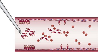
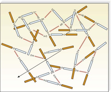
Detergent sclerosing solutions commonly used to treat varicose and telangiectatic veins include sodium morrhuate (SM), ethanolamine oleate (EO), STS, and POL (lauromacrogol 400, laureth-9). They produce endothelial damage through interference with cell surface lipids (Fig. 7.3). Strong detergents, such as STS and SM, produce maceration of the endothelium within 1 second of exposure.20 The intercellular ‘cement’ is disrupted, causing desquamation of endothelial cells in plaques. Because the hydrophilic and hydrophobic poles of the detergent molecule orient themselves so that the polar hydrophilic part is within the water and the hydrophobic part is away from the water, they appear as aggregates in solution (micelles) or fixed onto the endothelial surface (Fig. 7.4). Because the practitioner cannot ensure that the solution is entirely in contact with the endothelial surface (if the injected vein contains blood), the decrease in surface tension on the endothelial cells may not be in direct proportion to the concentration of the solution. Strong detergent sclerosants therefore have a low safety margin.20
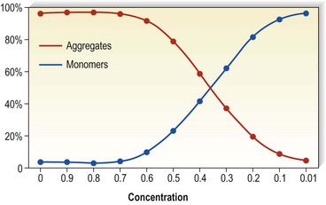
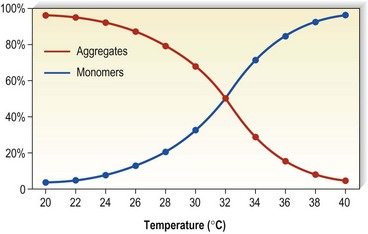
Detergents act as micelles when injected into a nondetergent environment (blood). Their destructive action on endothelial cells is enhanced when they act as aggregates rather than monomers. Thus, the concentration of the sclerosing solution in the vessel is an important factor regarding endothelial destruction and activity (Fig. 7.5). They have been found to aggregate to a significant extent at lower temperatures as opposed to room temperatures (Fig. 7.6).
Detergent sclerosing agents have been studied regarding their direct toxic effects on the formed elements of blood. One study found that the addition of SM, EO, STS, or POL to citrated plasma did not cause clotting nor shorten either the prothrombin time (PT) or the partial thromboplastin time (PTT).21 However, all of the sclerosing agents examined were directly toxic to both granulocytes and red blood cells (RBCs) at dilutions of up to 1 : 1000. When tested against cultured endothelial cells, all solutions were toxic to approximately 60% to 80% of cells at 1 : 100 dilutions, but only SM and EO were toxic at a further dilution of 1 : 1000. None of these tested solutions were toxic at 1 : 10,000 dilutions. Therefore, this study confirms that effective endosclerosis occurs through damage to endothelium and not through thrombosis induced by destruction or damage to red and/or white blood cells. Another in vitro study, however, found that the activated PTT was prolonged in proportion to the fall of factor XII and prekallikrein activity when POL was added to citrated serum.22 This indicates that in addition to its action on endothelial cells, POL is capable of acting on blood coagulation through activation of the early phase of the intrinsic pathway. The clinical relevance of this finding is unclear because additional studies have failed to demonstrate its significance, as explained later in this Chapter.23,24
Osmotic solutions
Hypertonic solutions, such as hypertonic saline (HS), probably cause dehydration of endothelial cells through osmosis, causing endothelial destruction (Fig. 7.7).22 It is speculated that fibrin deposition with thrombus formation on the damaged vessel wall occurs through modification of the electrostatic charge of the endothelial cells.20 For the vessel wall to be completely destroyed, the osmotic solution must be of sufficient concentration to diffuse throughout the entire vein wall.25 In contrast to the immediate action of detergent sclerosing solutions, experimental studies have shown that endothelial destruction with HS 22% or glucose 66% occurs only after 3 minutes.20 The destroyed endothelial cells do not appear to be desquamated as with detergent sclerosing solutions.20
Hypertonic solutions have a predictable destructive power that is proportional to their osmotic concentration. This was demonstrated in a comparative study of multiple hypertonic solutions used on the superficial and internal saphenous veins of 27 dogs.26 The degree of endothelial damage was assessed histologically at multiple times from 30 minutes to 8 weeks after sclerotherapy. The authors ranked the solutions from strongest to weakest as follows:
The authors concluded that maximal endothelial destruction occurred as early as 30 minutes to 4 days after injection, after which time the injected vessel went through either a reparative or a fibrotic process. Because dilution occurs with intravascular serum and blood, osmotic solutions have their greatest effect at or near the site of injection. In contrast, detergent sclerosing solutions can exert effective sclerosis for 5 to 10 cm along the course of the injected vessel. Sadick27 examined the sclerosing effect of HS 23.4% and POL 0.5%. He found equal sclerosing effect (length) for these two solutions injected in a similar type of vein using identical techniques. Unfortunately, HS 23.4% is more potent (about two to three times more) than POL 0.5%. Therefore, he inadvertently demonstrated that detergent solutions have about twice the therapeutic efficacy of osmotic solutions. A better comparison would have been with HS 11.7%.
Chemical solutions
Chemical irritants also act directly on endothelial cells to produce endosclerosis. Lindemayr and Santler28 studied the sclerosing effect of 4% polyiodinated iodine (PII; Variglobin) with standard and immunofluorescent microscopy and demonstrated fibrin deposition on the sclerosed veins only. Platelets fixed only to elastin, collagen, the basement membrane, and the amorphous material of the subendothelial layer, not to intact endothelial cells. It is also thought that the chemical destruction is in part related to the dissolution of intercellular cement, which has been demonstrated to occur after 30 seconds of exposure.20 Thus, this chemical irritant sclerosing solution produces its end result of vascular fibrosis through the irreversible destruction of endothelial cells with resultant thrombus formation on the subendothelial layer (Fig. 7.8).
The aforementioned mechanism of action has been demonstrated visually with scanning electron microscopy of sclerosed rabbit veins (agent not noted) by Merlen.29 He demonstrated intimal cracks and fissures that left intimal connective tissue fibers and elastic lamina exposed. Ultrastructural damage involving stasis of blood and platelet aggregation on intact endothelial intima occurred in 5 minutes in the dorsal rabbit ear vein.
Factors Predisposing to Thrombosis
As discussed previously, optimal clinical results occur when sclerotherapy-induced thrombosis is minimized. Factors predisposing to thrombus formation include decreased velocity of blood flow, hypercoagulability, and endothelial cell damage.30 The velocity of blood flow is unaffected by the sclerosing agent itself, but flow in general is usually slower in varicose veins and telangiectasia. This relative decrease in blood flow may predispose to thrombus formation in varicose veins and may be a significant contributing factor to the increased incidence of thrombophlebitis and deep vein thrombosis (DVT) in patients with varicose veins (see Chapter 2).
Hypercoagulability predisposes the patient to thrombus formation. Wuppermann31 studied fibrinolysis in subjects injected with POL and found a slight, statistically insignificant hyperfibrinolysis in blood drawn from the antecubital vein. He hypothesized that because coagulation factors II, VII, VIII, and IX and platelet function are damaged directly by the sclerosing solution, coagulation at the injection site is delayed. Endosclerosis, as measured by fibrinogen, gradually occurred over 5 days only at the injection site and did not result in systemic hypercoagulability. Therefore, sclerotherapy should not cause a sudden thrombosis. In fact, a hemolytic effect was detected with STS even at a 0.1% concentration, with POL at a 0.05% dilution, and with PII at a 2% concentration.23 The PTT, PT, and thrombin time (TT) were unchanged with injection of these agents into whole blood. Earlier work also demonstrated the lack of effect of POL on coagulation parameters in rabbits.32 MacGowen et al33 combined STS with whole normal blood, causing a homogeneous RBC lysate without the formation of thrombin. In vivo studies have also demonstrated a lack of hypercoagulability from sclerosing solutions. Cepelak24 found that platelet aggregation occurs only at the site of the sclerosing solution injection, with aggregation-inhibiting effects occurring in the efferent deep veins distal to the femoral vein. Thus, endothelial damage probably causes a release of various factors that produce anticoagulant effects, lowering the risk of thrombotic complications of sclerosing therapy. This effect was confirmed by Raymond-Martimbeau and Leclerc,34 who measured fibrinopeptide A and fibrin degradation of D-dimer fragments after injection of sodium iodine. They found no evidence for activation of blood coagulation. Therefore, experimental findings do not support the theory of intrinsic hypercoagulability of sclerosing solutions as the mechanism of action for thrombus formation during sclerotherapy (see Chapter 1).
The lack of hypercoagulability just mentioned correlates with clinical experience using POL. When POL is used in patients who are taking systemic anticoagulants, a decrease in its sclerosing action does not occur.35 The addition of heparin to STS also had no effect on the sclerotherapy results in a paired comparison of 100 patients.36 Thus it appears that the mechanism of action of POL and STS is to produce endothelial damage,37 but not thrombus formation associated with platelet aggregation. This mode of action is also seen with other (nondetergent) sclerosing agents. The low incidence of DVT after sclerotherapy (less than 1 per 10,000 sessions38) is the ultimate evidence that thrombosis is an epiphenomenon of the sclerosing process, not a goal.
Factors Predisposing to Endofibrosis
Whether vessels altered by changes of being varicose or stretched are more susceptible to the action of sclerosing agents than are normal vessels is unknown. At times, human varicose and telangiectatic vessels are noted to sclerose focally after the injection of various solutions. The focal nature of endothelial necrosis and thrombus formation may be related to toxic effects of the sclerosing solutions on the surrounding media. This effect is commonly observed when injecting varicose veins under duplex control. With this technique (described in Chapter 9), the sclerosing solution is injected and/or held in place until the varicosity is seen to spasm. This indicates effective sclerosis. Venograms of varicose veins injected with STS demonstrate segmental, intense, and diffuse spasm, both proximally and distally, at the time of injection and 6 minutes after injection.39 Effective endosclerosis occurs at points of vessel spasm where the entire endothelium is adherent. This agrees with the clinical impression that total compression of the sclerosed vessel is necessary for ideal, long-lasting, and complication-free sclerosis.40,41
At one time it was thought that ‘any solution which will not produce a slough when injected perivenously will generally not be strong enough to obliterate a vein.’42 However, some very effective sclerosing solutions are thought to act selectively on ‘damaged’ varicose endothelium. In fact, experimental studies have documented that effective sclerosing solutions do not have to produce tissue necrosis on intradermal injection (see Chapter 8). The manufacturers of POL state that this agent acts selectively on damaged vessels.a In addition, a number of histologic studies of the effect of sclerotherapy on varicose veins have also concluded that damaged varices are preferentially sclerosed.b,8,20,29 However, experimental injection of sclerosing agents into normal dorsal veins of rabbit ears yields effective vessel sclerosis in a concentration-dependent manner.43,44 Therefore, in addition to the type and concentration of sclerosing solution, other factors, including vessel diameter, rate of blood flow, and anatomic site of the vessel, may also be important.
Sclerosing solutions affect arteries in a different manner than they do veins. Although thrombosis occurs, intimal damage may not. MacGowen et al33 studied the local effects of intra-arterial injection of STS. They injected STS 3.0% into the central auricular artery at the base of the rabbit ear and visualized the resulting chain of events through a Perspex ear chamber (Lucite International, Southampton, UK) with high-power and oil-immersion lenses. Spasm was not noted in any vessels, but within minutes the erythrocytes appeared distorted and broken, with the formation of a central homogeneous thrombus that moved down the arteriole and lodged in a capillary. Intimal damage did not occur. Thus, the major effect of STS was on the blood cell mass that it destroyed and converted into an intravascular embolus. Subsequent biopsies of the ears at 1 hour and at 5 days demonstrated thrombus only, without evidence of intimal damage. These results are distinctly contrary to the effects of sclerosis on veins.
The reason for the different mechanism of action is unknown but may relate to the difference in velocity of blood flow in arteries and veins. Specifically, STS injected intra-arterially may not have enough time to react with endothelium, being both absorbed and inactivated by formed elements in the blood and serum factors and thereby being diluted to a ‘safe’ concentration by the more rapid arterial flow. Safe is a relative term, since inadvertent intra-arterial injections of STS have produced gangrene through thrombosis of vessels downstream of injection (see Chapter 8).
Experimental Evaluation of Sclerosing Solutions
An important question regarding the studies in this section is whether the experimental animal model is an appropriate system in which to compare the efficacy of various sclerosing solutions. The dorsal marginal rabbit ear vein is similar in size (0.35 to 0.45 mm in diameter) to telangiectasias in humans. Reiner45 found that it was difficult to measure the rate of dilution of sclerosing solutions in the rabbit ear vein because of the greater number of collaterals and rapid blood flow caused by the thermoregulatory nature of the ear. Therefore, after injection of the solution, 20 seconds of occlusion on the proximal and distal aspects of the injected vein wall caused by firm pressure helped simulate the more sluggish blood flow of human telangiectasias.43,44 In vitro studies of the effect of sclerosing solution on saphenous veins harvested for coronary bypass surgery have confirmed the histologic effect of sclerosing solution type and concentration with the rabbit ear vein model.46 However, study of an animal model may not produce accurate data because the researcher is comparing the action of a sclerosing solution on a normal vessel. In addition, the injected vessels are not compressed in rabbit ear vein studies, thereby resulting in the formation of a larger thrombus, which may allow for a more rapid or increased incidence of recanalization. Finally, thrombogenesis and thrombolytic effects are different in the rabbit ear than in the human artery and vein.47 However, despite all these shortcomings, as a model the rabbit ear vein does allow the physician to compare the mechanism of action of various sclerosing solutions both clinically and histologically. On the basis of these studies, the physician can achieve a similar therapeutic effect in humans by varying the concentration and type of solution (Table 7.2).
Table 7.2 Relative potency of sclerosing solutions
| Vein Diameter | Sclerosing Solution |
|---|---|
| 0.4–1 mm | Glycerin 70% |
| Chromated glycerin 50% | |
| Polidocanol 0.25%–0.5% | |
| Sodium tetradecyl sulfate 0.1% | |
| Hypertonic saline 11.7% | |
| Sclerodex | |
| Ethanolamine oleate 2% | |
| Polyiodinated iodine 0.1% | |
| Sodium morrhuate 0.25%–0.5% | |
| 1–3 mm | Sodium tetradecyl sulfate 0.25% foam |
| Polidocanol 0.5% foam | |
| Polyiodinated iodine 0.5%–1.0% | |
| Ethanolamine oleate 5% | |
| Hypertonic saline 23.4% | |
| Sodium morrhuate 1.0%–2.5% | |
| 3–5 mm | Polidocanol 1% foam |
| Sodium tetradecyl sulfate 0.5%–1.0% foam | |
| Sodium morrhuate 5% | |
| Polyiodinated iodine 2% | |
| >5 mm | Polidocanol 3%–4% foam |
| Perforators | Sodium tetradecyl sulfate 2%–3% foam |
| Saphenofemoral/popliteal junctions | Polyiodinated iodine 3%–12% |
In the 1920s, the first studies to elucidate the mechanism of action of sclerosing agents were performed using the dorsal vein of the rabbit ear. Sclerosing agents tested included 1% bichloride of mercury,48 30% sodium chloride and 50% grape sugar,49,50 30% sodium salicylate,51 50% to 60% calorose,52,53 and SM.54,55 All of these solutions achieved venous obliteration through endothelial cell alteration, with inflammation resulting in thrombus formation and the eventual production of a fibrous cord.
An evaluation of foamed sclerosing solutions has also been performed in vivo on isolated saphenous veins before stripping.56 Pathologic damage from 3% STS foam prepared with 1 mL of STS and 4 mL of air was extremely rapid, with complete damage to the endothelium within 2 minutes. Edema of the intima with progressive separation from the tunica media and formation of thrombus occurred at 15 and 30 minutes.
Sodium tetradecyl sulfate
The mechanism of sclerosis for intravascular STS was elucidated by Schneider8 and by Schneider and Fischer55 in human varicose veins, by Dietrich and Sinapius57 in rabbit external jugular veins, and by Imhoff and Stemmer20 in the dorsal rabbit ear vein. With STS, endothelial damage is dependent on concentration and occurs immediately after injection, with resulting rapid thrombus formation leading to vascular sclerosis.
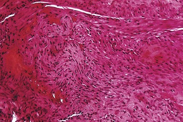
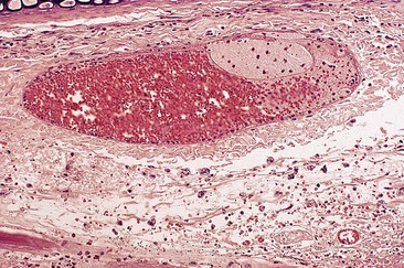
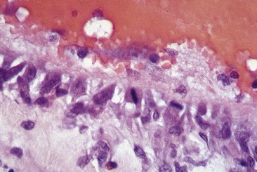
In two studies, sclerosis with STS produced similar results in a concentration-dependent manner.43,44 Endothelial damage occurred within 1 hour (Fig. 7.9), followed by the rapid onset of vascular thrombosis with subsequent organization (Fig. 7.10). Histologic recanalization occurred after 30 days with solution concentrations of 0.1% to 0.5%. The histologic findings explained the clinical appearance, which demonstrated initial thrombosis, followed by partial reappearance of the vessel injected with STS 0.1%. Therefore, there may be a concentration gradient in which an ideal concentration depends on many factors, including vessel diameter, rate of blood flow, animal model, and anatomic region within each animal model.
Sodium morrhuate
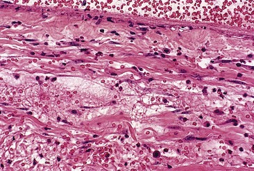
Sodium morrhuate, a mixture of sodium salts of the saturated and unsaturated fatty acids present in cod-liver oil, has been studied in the rabbit ear vein model.58 A 0.5% concentration of SM produced no clinical evidence of endothelial damage. Temporary histologic evidence of thrombosis was noted at 1 hour only, with a mild perivascular mixed cellular infiltrate (MCI). There was no evidence for extravasation of RBCs. Vessels injected with SM 1.0% were thrombosed between 2 and 10 days, after which the vessels normalized. Histologically, the SM 1.0%-injected vessel demonstrated a partially destroyed endothelium with extravasation of RBCs. The vessels injected with SM 2.5% demonstrated clinical fibrosis with histologic evidence of microangiopathic recanalization through a fibrotic cord at 45 days after injection. A unique finding noted with injection of SM, both 1.0% and 2.5%, was the presence of large numbers of perivascular mast cells (Fig. 7.11). This finding may correlate with the increased inflammatory nature and allergenicity of SM as compared with other sclerosing solutions.
Ethanolamine oleate
A synthetic mixture of ethanolamine and oleic acid, EO is another sclerosing solution that has been studied in the rabbit ear vein model.58 No histologic or clinical changes were noted with injection of EO 0.5%. Although an organizing thrombus was produced, complete recanalization occurred in the vessel injected with EO 1%, causing the returned clinical appearance of the injected vessel. Vessels injected with EO 2.5% had a partially destroyed endothelium followed by luminal recanalization. Evidence of phagocytosis of lipid-like material was noted in a vessel injected with EO 2.5% at 48 hours (Fig. 7.12). This may indicate extravasation of sclerosing solution either during injection or with endothelial destruction. Large numbers of perivascular mast cells were also noted 2 days after injection with EO 1% and 2.5%. Extravasated RBCs occurred in vessels injected with EO 1% and 2.5% at 1 hour and 2 days, but not in vessels injected with EO 0.5%.
A previous study comparing EO with STS was performed using the rat tail vein model.59 In this model, EO 5% was compared with STS 3% and 1%. Solution measuring 0.1 ml was injected and the veins were biopsied at 4 weeks. In this study, EO 5% was effective in sclerosing only 25% of the treated veins, as opposed to a 73% efficacy with STS 1% and a near 100% efficacy with STS 3%. Therefore, results in the rabbit ear vein compare well with those in the rat tail vein.
Polidocanol
A concentration gradient was also demonstrated in a study of POL in concentrations of 0.25%, 0.5%, and 1.0%.43 Only vessels injected with POL 0.5% and 1% were clinically sclerosed, and only the vessels injected with POL 1% maintained sclerosis without revascularization by 60 days.
An examination of the histologic effects of POL on the endothelium specifically, between 1 hour and 4 days after injection, illustrates the effect of varying the concentration of sclerosing solutions. Endothelial cells exposed to POL 0.25% were at first only partially damaged (Fig. 7.13). Mitotic figures indicating endothelial regeneration were noted 4 days after injection (Fig. 7.14). Likewise, with POL 0.5%, partial luminal recanalization occurred through an initial fibrotic cord in vessels (Fig. 7.15). Only vessels sclerosed with POL 1.0% developed complete endosclerosis (Fig. 7.16). Therefore, POL is probably a weaker detergent type of sclerosing solution than STS, and higher concentrations are necessary to produce complete vascular sclerosis.

Polidocanol: liquid versus foam
Hamel-Desnos et al and Wollmann have demonstrated both clinical60,61 and microscopic62 superior efficacy of foam versus liquid. In vitro studies have been conducted on single layers of endothelial cells in contact for 1.5 seconds with various concentrations of foam and liquid POL. Histologic examination demonstrated identical cell destruction with 0.5% foam and 3% liquid. As indicated by Hamel-Desnos et al, better efficacy had been supposed to be related to a longer time of contact; they have observed that the effect happened in a very short time, therefore there should be missing explanatory links. From a macroscopic point of view, foam seems to work because of the delayed dilution and closer contact between nondiluted sclerosing agent and endothelium; some microscopic phenomena will perhaps be found explaining more precisely what happens. (Foam sclerosants are described in detail at the end of this chapter and in Chapter 9.)
Hypertonic saline
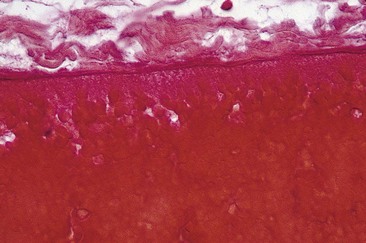
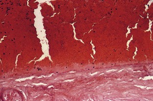
The sclerosing effect of HS was examined histologically in the external jugular vein of the dog by Kern and Angle,63 and in human varicose veins by McPheeters and Anderson.53 These investigators noted endothelial damage with thrombus formation within 1 hour of injection, with ultimate conversion into a fibrous cord within 2 to 4 weeks. This was confirmed in the rabbit ear vein model43 both clinically and histologically. Examination of the marginal ear vein 1 hour after exposure to HS 23.4% demonstrated complete endothelial destruction (Fig. 7.17).
However, subsequent evaluation of HS 11.7% in the rabbit ear vein model58 demonstrated an immediate thrombosis that lasted only 48 hours before complete normalization. Endothelial destruction was patchy at 1 hour with perivascular and intraluminal margination of polymorphonuclear cells and eosinophils. There was no evidence of extravasation of RBCs in the veins injected with HS 11.7%, whereas extravasation was noted in 30% of vessels injected with HS 23.4%. Therefore, the degree of endothelial damage and resulting extravasation of RBCs is proportional to the concentration of HS used.
Hypertonic glucose/saline
Sclerodex (SX; Omega Laboratories, Montreal) is a mixture of dextrose, sodium chloride, propylene glycol, and phenethyl alcohol. When SX was studied in the rabbit ear vein model,58 it produced an immediate thrombosis that lasted for 2 days, after which the vessel recanalized. At 1 hour, perivascular and intraluminal margination of polymorphonuclear cells and eosinophils were present with patchy endothelial destruction. Endothelial mitoses were present at 2 days within a regenerative endothelium. Extravasation of RBCs was not noted. Therefore, SX has a potency similar to HS 11.7%.
Chromated glycerin and 72% glycerin
The effect of chemical irritant sclerosing solutions has been studied in the rabbit ear model.44 Chromated glycerin (CG; Sclérémo, Laboratoires Bailleul, Paris, France), 50% and 100%, was injected into the dorsal marginal rabbit ear vein, producing clinical and histologic thrombosis that lasted only 2 to 8 days, after which the vessel appeared clinically and histologically normal. As noted with POL 0.25% above, the endothelium 1 hour after biopsy was almost undamaged. Therefore, in this experimental model, CG is a weak solution with a sclerosing effect similar to POL 0.25%. This correlates well with its clinical profile. The sclerosing effect of CG is not dependent upon chromium. Excellent results can be achieved in treating leg veins less than 1 mm in diameter with the use of a 72% glycerin solution mixed 2 : 1 with 1% lidocaine with epinephrine, as described later in this chapter.
Polyiodinated iodine
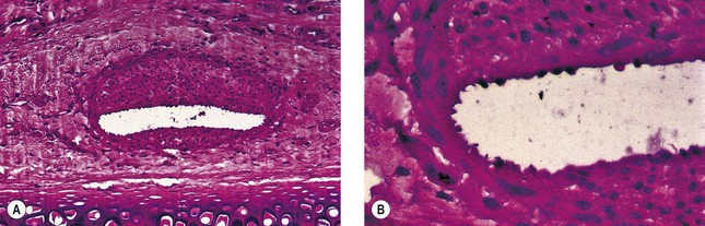
Various iodine solutions, with or without hypertonic solutions, have been examined in the rabbit ear vein model.64 Sclerodine (Omega Laboratories, Montreal) is a mixture of iodine USP (60 mg/mL) and sodium iodide USP (90 mg/mL). This solution was compared with an American iodine formula consisting of iodine and sodium iodide, compounded by the Women’s Hospital of Texas in Houston. The two sclerosants were identical in composition. After being mixed with either normal saline, SX (Omega Laboratories), or a solution of dextrose 250 mg/mL and sodium chloride 100 mg/mL (compounded by the Women’s Hospital of Texas in Houston), each sclerosant was injected in concentrations of 0.1% and 0.5%. Thrombosis occurred with all solutions at 1 hour, with an attenuated endothelium and focal endothelial necrosis. A mild perivascular mixed cellular infiltrate consisting of eosinophils and polymorphonucleocytes was present with margination along the endothelial border. With the 0.1% solutions, the endothelium was hyperplastic, with regenerative changes noted by 8 days (Fig. 7.18) and complete normalization by 28 days. With the 0.5% solutions, the endothelium was necrotic with multiple brown spherules approximately 1 µm in diameter seen within the endothelial wall (Fig. 7.19). These may represent iodine crystals. By 28 days, a fibrous cord was present without inflammation. Interestingly, veins treated with iodine/normal saline mix showed a greater incidence and extent of extravasated erythrocytes compared to those treated with iodine/SX (hypertonic saline/dextrose) solutions. The PII 0.1% solution had an experimental efficacy equivalent to HS 11.7%, SM 2.5%, STS 0.25%, and POL 0.5%. The PII 0.5% solution had an experimental efficacy similar to HS 23.4%, STS 0.5%, and POL 1.0%.
< div class='tao-gold-member'>
Related posts:

Stay updated, free articles. Join our Telegram channel

Full access? Get Clinical Tree


