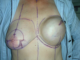
Fig. 61.1
(a, b) Preoperative view. A 51-year-old patient had a prior quadrantectomy and radiation for an invasive cancer in the upper outer quadrant of the left breast with the scar extending into the axilla. There was a local cancer recurrence in the periareolar region which was diagnosed through an open biopsy. Both breasts were of medium size with moderate ptosis with the left breast being smaller compared to the right breast and the nipple dislocated toward the axilla
61.2 Surgery
A skin-sparing mastectomy with resection of the nipple-areola complex was performed with both scars included in the mastectomy incisions. The patient had a prior axillary lymph node dissection, and reoperation sentinel lymph node biopsy found that no sentinel nodes were found intraoperatively neither with the blue dye nor in the preoperative scintigraphy. A submuscular pocket was dissected under the major pectoralis muscle, and serratus anterior muscle and a 450 cc anatomical expander was inserted and filled with 100 cc of sterile saline solution. After completion of wound healing, the expander was filled every 2 weeks with 100 cc saline solution up to a total filling volume of 400 cc. Following 6 months of expansion, the expander was exchanged to a 295 cc anatomical silicone implant. Concomitantly a reduction mammoplasty was done on the right breast for symmetrization (Fig. 61.2).


