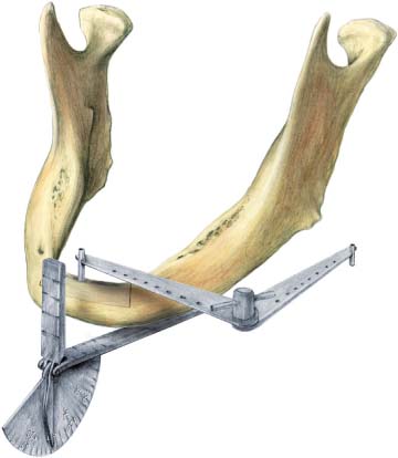
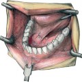
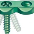
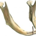
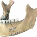
32 Mandible Alveolar Bone Reconstruction by Endodistraction Traditionally, for functional rehabilitation of severely atrophic edentulous mandibles, short implants with long abutments are used. Another possibility for treatment is bony reconstruction by bone transplantation. Recently, distraction osteogenesis has offered a third option for reconstruction of the alveolar process. The simple principle is based on the natural healing process of fractured or split bones to grow together by osteogenesis, and is called callus distraction (Ilizarov, 1989). Other common distraction appliances are fixed to the bones with two plates, and there is a need for bulky rods projecting outward into the oral cavity. The endodistraction screw sits in a tap-hole in the body of the mandible and is combined with a hollow implant and tap, which is anchored to the osteotomized superior segment of the bone and rests on a metal shoulder of the endodistraction screw. The endodistraction screw appears up to 10 mm beyond the inferior border of the mandible, hidden in the soft tissues of the chin. This distance corresponds to the possible amount of distraction osteogenesis. By turning the endodistraction screw, the hollow implant together with the osteotomized bone is lifted up step by step; the osteotomy gap increases, and distraction osteogenesis begins. The main indication for this technique is a mandible classified as Cawood 5 or 6 (Cawood and Howell, 1988). The best anatomical predisposition for distraction osteo-genesis is in the anterior aspect of the mandible, because of its good blood supply from the Tsusaki vessels on the lingual side (Tsusaki, 1955), which are terminal branches of the sublingual arteries (Krenkel et al., 1985). Fig. 32.1 Drilling in the right direction for the planned vector of distraction osteogenesis is performed with a Josef calibrator (Padgett, Germany, Nr. P690). The endodistraction method for vertical callus distraction osteogenesis is a single device, which allows callus formation to be realized in every desired height, width, and direction. For planning the correct vector of the distraction osteogenesis, we use lateral transcranial X-ray and simple drawings according to the ideal position of the intended dentures. Our reference plane is the inferior border of the mandible, which is reliable also during the operation. With the help of an angle-measuring device according to Josef (Fig. 32.1) during the operation, we choose the right angulation for the tap-hole of the endo-distraction screw in the basal arch. The marking is then performed, exactly in the midline of the alveolar crest. First a 2-mm diameter hole is drilled in the desired direction, through and beyond the basal arch. Then supraperiosteal dissection is performed from the vestibule of the mouth, and the mental nerves are identified. This is followed by the planning and marking of the osteotomy. A hole is traced and drilled in the edges of the osteotomy. The osteotomy itself can be performed either by a sagittal microsaw or by piezosurgery with ultrasound (Fig. 32.2
Introduction
Materials and Method
![]()
Stay updated, free articles. Join our Telegram channel

Full access? Get Clinical Tree


