Congenital
Aganglionosis (Hirschsprung’s disease)
Anorectal malformations
Acquired
Neuronal degeneration (Chagas’ disease)
Obstructive
Endocrine
Central nervous system
Psychiatric
Idiopathic
In the absence of an underlying organic cause, dilatation of the rectum is termed idiopathic megarectum (IMR) [13]. It is the management of patients with this condition that has proved most challenging.
Putative Etiology of Idiopathic Megarectum
The intriguing question about why visceral dilatation occurs in some functional disorders of the rectum or indeed anywhere in the gastrointestinal tract has not yet been answered. The pathogenic mechanism(s) and etiology of IMR are unknown, although neurophysiological, behavioral, and psychological abnormalities have all been implicated [5]. It is possible that more than one mechanism may be relevant in the development of IMR, even in individual cases. Broadly, two theories of the development of IMR have been proposed. The first is that IMR occurs secondary to a behavioral problem of defecation [14, 15] associated with a failure of toilet training, voluntary withholding of stool, or both, leading to rectal distension and ultimately loss of rectal contractility, impaired rectal function [5, 16], and, perhaps subsequently, secondary pathological abnormalities. The counter argument is that the primary abnormality lies within the rectal wall itself [17]. In such cases, an intrinsic neuromuscular disorder of the rectum (e.g., loss of rectal sensation [i.e. rectal hyposensitivity], or diminished contractility) may allow chronic accumulation of feces and rectal dilatation [17–19]. The etiologies of such primary dysfunction have rarely been addressed, and it is unclear whether the changes demonstrated are primary or secondary or inherently epiphenomena [20].
Pathology of Idiopathic Megarectum
IMR is a disorder that traverses the boundary of functional and organic gastrointestinal disease. That is, although there is clearly a disorder of function and attendant macroscopic disease (visceral dilatation), there is no universal histopathological abnormality that is diagnostic. Histopathological abnormalities of all three final effectors (the enteric nerve system, smooth muscle, and the interstitial cells of Cajal [ICCs]) and of sensorimotor function have been reported in IMR, and some generalizations can be drawn from a review of the available literature [20]. Studies of neuronal changes have revealed that morphological abnormalities are largely absent on routine and silver stains [12, 21–23], but variable changes in neurochemistry have been reported [24]. Studies of smooth muscle mostly report hypertrophy plus some degree of fibrosis. There have been no studies of ICCs specific to patients with IMR, but two studies of patients with megacolon have been performed, both of which report decreased density of ICCs [25, 26] and one of which demonstrated decreased process length [25]. In this respect, caution should be exercised in the interpretation of such findings because these observations do not necessarily prove disease causation [20, 27].
Clinical Approach to Patients with Megarectum
Patients with IMR suffer with symptoms of severe intractable constipation. It is useful, therefore, to consider the clinical approach to patients with IMR in the context of the management of patients presenting with chronic constipation, including how to identify the subgroup with dilated bowel. Comprehensive clinical evaluation of patients, with integration of the findings from clinical history, physical examination, and investigations, aims to achieve each of the following:
1.
To establish the severity and impact of clinical symptoms
2.
To identify constipated patients with rectal dilatation, allowing distinction from patients with normal rectal caliber
3.
To exclude coexisting pathology/underlying causes of megarectum, most notably adult HSCR
4.
To attempt to deduce the importance of putative etiological processes (neurophysiological versus psychobehavioral)
5.
To ensure that selection criteria for surgical intervention are met
Clinical Evaluation
A detailed and careful clinical history will reveal important information relating to several key areas: (1) severity of symptoms of constipation, (2) impact of symptoms on quality of life, (3) presence of associated symptoms such as soiling/fecal incontinence, and (4) possible etiology. An assessment of the severity of symptoms may be made by recording details related to the frequency (or, more accurately, the infrequency) of defectation and the effectiveness of evacuatory function. It is important to ascertain the onset and duration of symptoms because those with onset of symptoms during childhood or adolescence invariably experience recurrent fecal impaction with soiling, whereas patients with a “late onset” of symptoms tend not to complain of this symptom [6, 28] Additional relevant information includes the usual stool consistency and the percentage of defecatory attempts that are associated with (1) the need to strain, (2) a sensation of anorectal obstruction or incomplete emptying, and (3) defecation attempts that are ultimately unsuccessful. The usual time required to defaecate and the need for digitation (either vaginally or anally) to facilitate rectal emptying should be noted. Abdominal pain, distension, and regular use of a laxative/suppository/enema are present in the vast majority of patients [6, 22, 28], and their extent and specific nature also should be documented.
It is advisable to obtain objective information about a patient’s symptoms and their severity using validated constipation (with or without fecal incontinence; discussed later) scoring systems (such as the Cleveland Clinic constipation score) [29]. In a research setting, the Rome III diagnostic criteria for chronic constipation [30] should be sought and documented. It is also important to gauge the impact of symptoms on the patient’s quality of life by assessing their effect on activities of daily living and sociopsychological status, which can be formalized using the patient assessment of constipation–quality of life questionnaire [31].
For the purposes of this chapter, it is assumed that other possible organic lower gastrointestinal diseases have already been excluded by appropriate assessment. A full enquiry about all lower gastrointestinal symptoms is clearly important in facilitating this. In addition, in the context of megarectum, it is vital to enquire about soiling and fecal incontinence and, when present, to record detailed information relating to the frequency and amount of leakage of solid and liquid stool and flatus. Again, the use of a validated scoring system (such as the Cleveland Clinic score [32]) provides objective information concerning the severity. Symptoms of pelvic organ and rectal prolapse must be sought.
A detailed history also provides the opportunity for the clinician to exclude other causes of megarectum, for example, psychiatric, endocrine, or central nervous system diseases, including possible underlying psychobehavioral issues (e.g., functional fecal retention during childhood, toilet avoidance during adulthood, or both). A detailed interview with the patient also provides the opportunity to ensure that all nonsurgical interventions have been exhausted.
A thorough general and abdominal examination should be performed. In patients with gross dilatation and fecal impaction of the rectum (especially when associated with concomitant colonic dilatation), it may be possible to palpate a large fecaloma during abdominal examination. This can be recognized as a mass arising from the pelvis that “indents” on palpation. Of particular relevance to the assessment of patients with IMR is the examination of the neurological system and the anorectum. With respect to neurological evaluation, attention should be focused on somatic lumbosacral spinal cord function; an assessment of gait and neurological function of the lower limbs should be performed in all cases. If neurological disease is likely, more detailed examination is clearly warranted.
When examining the anorectum, the patient is usually positioned in the left lateral position with the hips flexed, although the jack-knife position also is used. The examination begins with inspection of the anus and perineum. Evidence of congenital anomalies should be noted. The perianal skin and surrounding area should be checked for the presence of fecal soiling, mucus or purulent discharge, and excoriation. Any skin tags, external hemorrhoids, fistulae, or other conditions should be noted and clearly documented. These findings are often present in patients with IMR because of chronic evacuatory difficulties and each may need to be addressed separately.
The anal canal is usually closed at rest; a patulous anus may suggest underlying sphincter dysfunction (see the section “Physiological Investigation of Patients with IMR”). The examination proceeds with digital examination of the anorectum. A basic idea of resting tone and strength of voluntary contraction can be gained, although this is not necessarily accurate. The rectum is examined for masses and the presence of impacted feces. A sense of the “capacity” of the rectum also can be appreciated during digital examination. The examination is completed by performing a proctosigmoidoscopy to exclude local organic pathology of the distal bowel and functional anatomical disorders (prolapsing hemorrhoids/rectal mucosa and rectocele). The finding of a solitary rectal ulcer may confirm the presence of an underlying evacuatory disorder. In our experience, rigid sigmoidoscopic examination also provides the surgeon with a useful assessment of the capacity/elongation of the rectum because the scope often “falls in” to its maximum length with significant ease in patients with IMR, provided, of course, the rectum is empty of impacted feces.
Although certain features of the patient’s history and physical examination may suggest the presence of the most extreme cases of megarectum (such as extreme infrequency of defecation or recurrent fecal impaction with soiling, large fecoloma on abdominal palpation, gross fecal impaction of the rectum on digital examination, obvious dilatation/elongation of the rectum during rigid sigmoidoscopic examination), because clinical features alone are unreliable in identifying such patients, it is likely that milder forms of the condition may remain unrecognized and thus untreated in patients presenting with constipation.
Investigation of Patients with Suspected Megarectum
From a clinical perspective, identification of patients with megarectum can be challenging. It is essential to appreciate, as alluded to earlier, that there is no “predictable” or reliable clinical presentation that will alert the clinician to the potential diagnosis. In part, this is because gastrointestinal symptoms alone are not specific and are insensitive for different disease processes and they do not provide an accurate indication of underlying pathology. This principle is all the more relevant in the assessment of functional gastrointestinal disorders, where it is increasingly appreciated that formal investigation is required to identify apparently homogenous subgroups, such as those with megarectum.
In this section about investigation, therefore, the following areas will be addressed:
1.
The investigative strategy to identify the subgroup of constipated patients with megarectum
2.
Confirmation of the diagnosis of megarectum/assessment of colonic caliber
3.
Establishment of a diagnosis of IMR by exclusion of other underlying causes of megarectum, including adult HSCR
4.
Consideration of novel investigations for further definition and subclassification of patients with IMR
Investigative Strategy to Identify the Subgroup of Constipated Patients with Megarectum
Clearly, it would be impossible and inappropriate to investigate all patients presenting with symptoms of chronic constipation, given that its prevalence approaches 10 % in Western populations [33]. Therefore, investigation of such patients needs to be rationalized and selective. In our practice, we recommend further investigation to those patients with severe, intractable, chronic constipation in whom there is a significant impact on quality of life and in whom simple measures to treat the constipation have failed to alleviate symptoms. This is somewhat biased toward patients referred to a tertiary referral center that specializes in the medical and surgical management of such cases. Assuming that underlying organic disease has been excluded, we undertake investigation in accordance with current recommendations [34]. Measurement of serum calcium levels and thyroid function tests are performed, accepting that the diagnostic yield of such tests will be low. Subsequently, constipated patients are subjected to rigorous laboratory assessment of colonic and anorectal function using comprehensive physiological investigation. We routinely perform the following physiological tests in all constipated patients who are referred for specialist investigation:
Anorectal manometry to assess anal sphincter function, rectal sensitivity (see below), and rectoanal reflexes [35]
Neurophysiological evaluation to assess pelvic floor innervation using pudendal nerve terminal motor latencies [35]
Ultrasonographic evaluation to assess the morphological integrity of the anal sphincter complex
Evacuation protography (some investigators use the balloon expulsion test) to assess rectal evacuatory function and to identify concomitant radiological abnormalities of the rectum (e.g., rectocoele/rectal intussusception)
Colonic transit studies to assess colonic transit time for the identification of slow colonic transit. Initially, we use radiopaque marker studies and colonic scintigraphy in proven cases of slow-transit constipation.
Magnetic resonance imaging (MRI) in selected cases to determine preliminary rectal volume and caliber as well as rectal emptying [36]
Findings of Physiological Investigation in Patients with IMR
Despite an unknown etiology, evaluation of anorectal function has revealed that patients with IMR have excessive laxity (increased compliance) [37–39] and sensory dysfunction [6, 38] of the rectum. Furthermore, patients also have impaired rectal evacuatory function [6, 37], often with secondary delay in colonic transit [39].
The results of anal manometry have frequently been reported in patients with IMR. In terms of anal canal pressures, reduced resting, but not squeeze, anal pressures have been noted [3, 6]. It has been suggested that these result from chronic relaxation secondary to the presence of a fecoloma [3] because sphincter function is recoverable after surgical correction of the dilated rectum [40]. However, it may also reflect internal anal sphincter damage sustained during manual disimpaction of the rectum under general anaesthesia [41], and thus endoanal ultrasonographic evaluation of the sphincter complex is advisable in these cases.
The abnormalities of sensorimotor function are well documented in patients with IMR and deserve special consideration. First, delayed colonic transit commonly coexists with IMR [38, 42, 43], occurring predominantly in the dilated segments of gut [3, 38]. Second, impaired perception of rectal distension, as evidenced by elevated sensory threshold volumes required to elicit rectal sensations during balloon distension, is also almost universal among patients with IMR [6, 42, 43].
Accordingly, the finding of impaired perception of rectal distension in constipated patients should alert the clinician to the possibility of the diagnosis of megarectum and should prompt more detailed evaluation (see the section “Confirmation of the Diagnosis of Megarectum”). It is important to note that although this finding of “rectal hyposensitivity” is universally present in patients with megarectum (when the rectum is empty of feces), it is not specific and can be present in patients with a normal rectal caliber on account of impaired afferent nerve function [44].
Confirmation of the Diagnosis of Megarectum/Assessment of Colonic Caliber
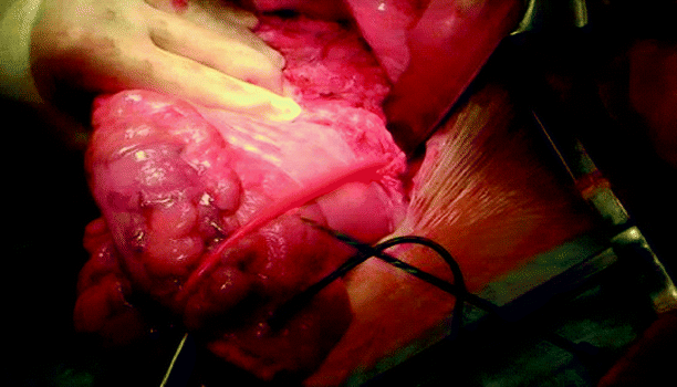
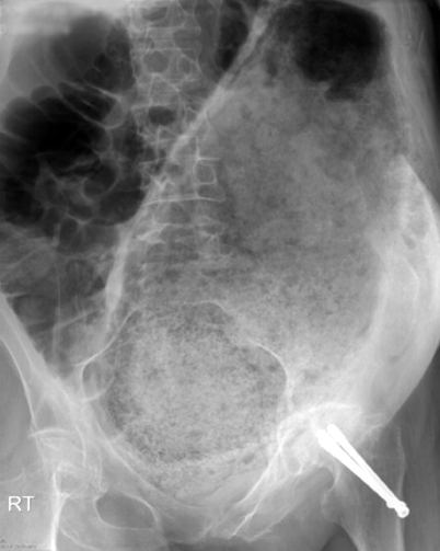
The term megarectum is often used indiscriminately. Dilatation of the rectum may be so gross that it is evident during clinical examination or laparotomy (Fig. 25.1) or on a plain radiograph of the abdomen (Fig. 25.2), although diagnosis on this basis is somewhat subjective because no specific criteria exist. Traditionally, two methods have been employed to make a diagnosis of megarectum.


Fig. 25.1
Example of megarectum encountered during laparotomy

Fig. 25.2
Plain abdominal radiograph of a patient with megarectum. The grossly dilated rectum is impacted with feces and can be seen extending out of the pelvis toward the left upper quadrant
First, the diagnosis has usually been made when the rectal diameter at the pelvic brim is greater than 6.5 cm on a lateral radiograph obtained during double-contrast barium enema [45]. In recent years, evacuation proctography, which also utilizes radiopaque contrast, has been used in the assessment of constipated patients, and although used, the above definition has not been validated for this technique. It is important to appreciate that contrast studies may not accurately reflect or assess rectal dimensions [46, 47] because they rely on volumetric distension and do not control for the differing rectal volumes or distensibilities seen in constipated patients [48, 49], which, coupled with proximal progression into the sigmoid and possible anal leakage of contrast, may result in incomplete distension and thus an overall underestimation of rectal diameter [8].
Alternatively, a diagnosis of megarectum has been made during anorectal manometry on the basis of an elevated maximum tolerable volume during simple “latex” balloon distension [37, 50], which is assumed to reflect rectal capacity [51–53]. However, an increase in tolerated rectal volume does not necessarily indicate an increased rectal diameter (and thus a diagnosis of megarectum) because it also is influenced by the sensitivity of the afferent nerves supplying the rectum [49] and thus will be elevated when there are nerve signaling problems along this pathway (such as pelvic nerve or spinal cord injury) [44]. In addition, axial expansion and rostral extension of rectal balloons into the sigmoid colon will influence the sensory threshold volumes [54]. Rostral extrusion of rectal balloons and assumptions regarding their intrinsic compliance affect compliance measurements of the rectum, as does the inherent rectal geometry, which is disturbed in megarectum: Newer technologies such as impedance planimetry and the barostat (discussed later) that equate the estimated cross-sectional area of the rectum during standardized rectal distension are needed to separate these cases [55, 56].
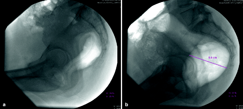
Given the fundamental methodological limitations of the above techniques, a novel technique for the diagnosis of megarectum has been developed by our group [8]. This technique involves the use of a barostat, which is a computerized device capable of achieving a constant distending pressure in the viscus under evaluation by changing the volume of air insufflated and which is now the method of choice for the evaluation of various components of rectal sensorimotor function [57–59]. Controlled isobaric distension of the rectum using a barostat during fluoroscopic screening allows rectal diameter to be assessed accurately and consistently. A diagnosis of megarectum was made when the maximum rectal diameter was >6.3 cm at the minimum distending pressure of the rectum (the pressure at which sufficient air is insufflated to prevent collapse of the wall but not cause active distension of the rectum) [8]. An example of a patient with megarectum compared with a constipated patient with a normal-caliber rectum is shown in Fig. 25.3a, b. This technique seems to be well tolerated, demonstrates excellent interobserver agreement, and has criteria for diagnosis defined by a normal range established in asymptomatic controls [8]. Furthermore, the use of a barostat to perform controlled distension to a specific, predefined “threshold” ensured standardization of methodology between subjects [8].


Fig. 25.3
Rectal diameters in constipated patients during rectal distension using a barostat. Fluoroscopic images of the rectum in patients with chronic constipation obtained during isobaric distension at minimum distending pressure reveal (a) normal diameter rectum and (b) megarectum
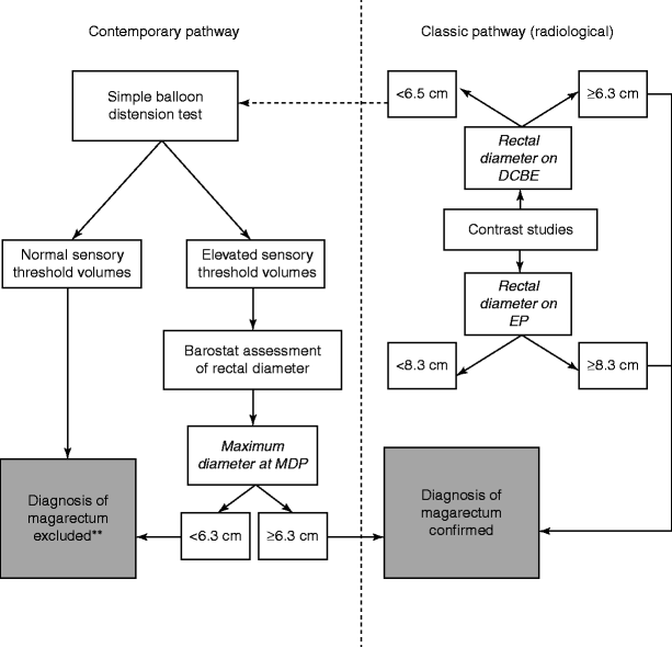
On the basis of all available diagnostic techniques, taking their inherent limitations into consideration, a contemporary strategy for the diagnosis of megarectum recently has been proposed (Fig. 25.4) [20]. In summary, the diagnosis of megarectum should be made using one of the following objective criteria [20]:


Fig. 25.4
Diagnostic algorithm for patients with suspected megarectum. The preferred option for the initial investigation of patients with suspected megarectum is a latex balloon distension test because it avoids exposure to ionizing radiation in those without the condition.** normal sensory threshold volumes exclude the presence of megarectum on simple balloon distension only if the rectum is empty of feces. DCBE double-contrast barium enema, EP evacuation proctography, MDP minimum distending pressure of the rectum (Reprinted with permission from Gladman and Knowles [20])
1.
A rectal diameter at the pelvic brim of greater than 6.5 cm on a lateral radiograph obtained during double-contrast barium enema
2.
Widest rectal diameter of greater than 8.3 cm on lateral radiography during evacuation proctography [8]
3.
A rectal diameter of greater than 6.3 cm at the minimum distension pressure during controlled (pressure-based) distension using a barostat during anorectal physiology combined with fluoroscopic imaging [8]
In terms of planning, the presence or absence of concomitant proximal colonic dilatation (megacolon) is crucial when deciding appropriate surgical strategy (if clinically indicated). Unfortunately, of the three imaging methods mentioned above, only a double-contrast barium enema study will enable exclusion of proximal bowel dilatation and is thus considered mandatory before undertaking surgical intervention. However, it should be noted that this technique is not always sensitive for diagnosing megarectum and can underestimate dilatation [8]. Therefore, if clinical suspicion is high, the other techniques should also be employed. In contrast, a diagnosis based on elevated sensory threshold volumes during anorectal manometry should not be used because it is not sufficiently specific and overdiagnoses the condition [8].
Establishment of a Diagnosis of IMR by Exclusion of Other Underlying Causes of Megarectum, Including Adult HSCR
As alluded to previously, other causes for the development of rectal dilatation (see Table 25.1) should be excluded by appropriate assessment. For the most part, the most significant condition to be excluded is adult HSCR.
Exclusion of Adult HSCR
The distinction between IMR and adult HSCR is an area of diagnostic difficulty. Although much is made of this in textbooks, in reality—at least in the developed world—adult HSCR is exceedingly rare, with only 300 cases described in the literature to date. Even the largest series (that of the Mayo Clinic and St. Mark’s Hospital) each document seeing only about one case per year from the 1950s onward [60, 61]. In Western practice, the vast majority of instances of classic HSCR, in which a variable portion of rectosigmoid is affected, is now diagnosed in neonates and infants (even in utero) [62]. It is thus the short- and ultrashort-segment disease for which an index of suspicion and careful investigation should be undertaken in adults. Clinically, the finding of a meticulously clean anus, in contrast to the gross soiling commonly observed in IMR, should alert to the suspicion of HSCR. In contrast to classic disease—which is evident on barium enema, by an absence of rectoanal inhibitory reflex (RAIR), and on suction rectal biopsy—the diagnosis of short- and ultrashort-segment disease is more problematic. In the former, the RAIR is absent but biopsies are usually definitive; however, in the latter, both may be unreliable, especially when considering that the anal transition zone, which is affected in isolation in ultrashort-segment disease, can normally be hypoganglionic [63].
A further problem arises when using the RAIR to distinguish these conditions. Even accepting the larger volumes of distension required to achieve relaxation [57, 58], the reflex is absent in 30–50 % patients with IMR after exclusion of HSCR on biopsy [3, 6, 64]. This may be due to (1) lack of adequate stimulation of the rectal wall by the balloon in a capacious rectum with altered pressure/volume characteristics [39], (2) chronic relaxation of the internal sphincter due to fecal impaction [3], or (3) previous damage to the internal sphincter during manual evacuation under general anesthesia [41]. In our experience, it usually is necessary to perform a full-thickness rectal biopsy to confirm aganglionosis in patients with an absent RAIR during anorectal manometry to confirm or refute the diagnosis of HSCR.
Stay updated, free articles. Join our Telegram channel

Full access? Get Clinical Tree


