Fig. 52.1
Excision of four skin pits with a lateral incision for drainage
In a recent study of 1,435 cases, the midline pits and secondary openings were simply cored out using skin trephines and the abscesses were cleaned out through the trephines, which were left unsutured. There was a 16.2 % recurrence rate with a mean follow-up of nearly 7 years [53].
The relatively high recurrence rates after simple excision of skin pits [22 28 52–55] is tempered by the ease and success [7 22] of a repeat procedure, which means that although one operation may cure less than 85 % of patients, a second finally cures more than 95 % of all cases. Open wounds are the other disadvantage of these procedures. However, because 50 % of the lateral wounds after a Bascom procedure were completely healed at 3 weeks, 99 % by 3 months, and all wounds were healed by 4 months [52], and because patients can care for themselves in the community, an open lateral wound does not greatly delay return to normal activities [22 53].
Wide Excision of the Abscess With or Without Midline Skin Closure
Wide excision of the abscess in the elective setting will result in the debilitating complications from this type of surgery, including unhealed midline wounds [10 56–58]. There is little information concerning their frequency of occurrence, although it may be as high as 2–7 % [59 60]. In this respect, because unhealed wounds occur mostly in the midline and pilonidal disease is caused by the environment created by the natal cleft, it is likely that there will be a higher risk of this complication and recurrent disease if long midline wounds, whether sutured or unsutured, are created, as happens after many wide excisional procedures. Surgery is the main cause of prolonged unhealed wounds and a major cause of morbidity. It should be noted, however, that spontaneous unhealed wounds in the natal cleft without previous surgery also can occur.
Systematic reviews [4 5] confirm the conclusions of many previous nonrandomized studies of wide excisional techniques, and although time to complete healing may be quicker after midline closure, recurrence rates can be as high as 22–41 % [14 15] compared with approximately 6 % with the simple lay-open nonexcisional procedures [42–46 48 61].
Off-Midline Skin Closure With and Without Wide Excision of the Abscess
Two systematic reviews [4 5] that included an analysis of six randomized, controlled trials (RCTs) [4] concluded that after excision of the abscess, off-midline rather than midline skin closure should become standard management [62]. The Karydakis off-midline flap procedure (Fig.52.2) is achieved by an elliptical asymmetrical wide excision of skin, abscess, and sinus, mobilizing the skin edge closest to the midline to produce a thick flap, which is then sutured to the side farthest from the midline. The deep surface of the flap is transfixed to the underlying sacrococcygeal fascia [21]. In a study of 7,471 cases from 1966 to 1990, 95 % of whom were followed up for 2–20 years, Karydakis [25] found that the mean time in hospital was 1–3 days. The majority healed rapidly, with an average time off work of 9 days, and 55 patients (1 %) developed recurrences.
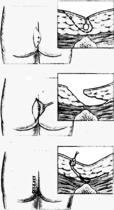
Fig. 52.2
Diagrammatic representation of the Karydakis procedure showing excision of the sinuses and abscess (top) together with the skin from one side of the natal cleft (middle) with off-center skin closure (bottom)
Kitchen’s [62 63] and Bascom’s [10 11 56] modifications of the Karydakis technique use thinner skin flaps, and the Bascom “cleft lift” procedure (Figs.52.3 52.4, and52.5) avoids any excision of the abscess or secondary openings. If the abscess wall is rigid, it is “cubed” to allow it to collapse. Secondary openings of the abscess well away from the midline do not need to be included in the excision wound. They will heal after the eradication of the primary midline skin pits if their openings are slightly enlarged for drainage and the tracks leading to them are cleaned of hairs and debris. Although initially the Karydakis procedures were done under general anaesthesia, the modified techniques increasingly are being performed under local anaesthesia as day case [11 62 63].
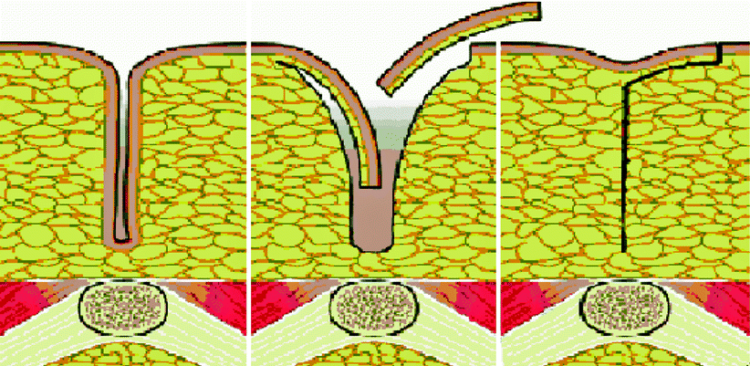
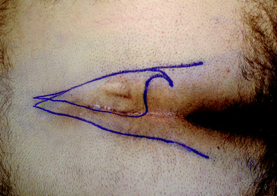
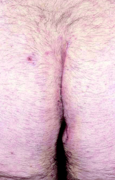
Fig. 52.3
Diagrammatic representation of Bascom’s cleft lift procedure showing excision of the skin from one side of the natal cleft (left, middle) with closure of the cleft and suture of the skin to one side of the midline (right)
Fig. 52.4
Outline of the skin for excision in Bascom’s cleft lift to include the midline skin pits and skin from the diseased side of the natal cleft. The outer lines mark where the skin edges meet naturally when the patient stands up
Fig. 52.5
A healed lateralized wound after a cleft lift with healthy skin in the midline
The number of publications about the Karydakis procedure [64–74] in the past few years demonstrates its increasing popularity, and most report similar good results apart from one RCT [75] that has shown a higher wound infection rate after this procedure compared with the Limberg flap (26 vs. 8 %). Although 4–26 % of patients have wound complications including skin necrosis, infections, seromas, hematomas, and wound dehiscence, most of these can be managed on an outpatient basis. Complete wound healing occurs within 1 week in 60–70 % of patients, of whom most require minimal care in the community and return to work within 1–4 weeks. The debilitating complications of surgery for pilonidal disease have not been reported after this technique.
A randomized trial of Bascom’s two operations in patients with primary disease showed better results with the cleft lift procedure compared with the simpler pit picking operation [76]. The cleft lift procedure may be the best operation for more extensive disease in hairy patients with deep natal clefts and multiple pits [76–80].
More Complex Skin Flap Procedures After Wide Excisional Surgery
Although good results have been achieved after Z/V-Y plasties and rhomboid/Limberg flaps [81–92], two RCTs suggested that V-Y and Limberg flap procedures were not superior to a simple primary closure technique [93] or laying open with marsupialization [94]. These more complex flap procedures all include excision of the abscess and require general anesthesia and a longer hospital stay, and they have occasional complications that need further inpatient surgery. Overall they offer little or no extra benefit to the modified Karydakis procedures and are cosmetically disfiguring [80].
Failures and Recurrences
In published work, it is often difficult to differentiate between failure and recurrence, and indeed, once a failure has continued for many months it may be seen by both the surgeon and the patient as being a recurrence. The management of these patients is part of the ongoing treatment of pilonidal sinus disease. The most debilitating consequence of failure is the unhealed midline wound.
Unhealed Midline Wounds After Surgical Treatment
Although unhealed midline wounds may be uncommon (Fig.52.6), they are thebête noire of pilonidal surgery, which, together with recurrent disease, the numbers of postoperative wound complications, and the time needed to complete healing of open or dehisced sutured wounds, are the factors that need to be measured to compare different modalities of treatment. Few studies have recorded all of these parameters [4], particularly the frequency of unhealed midline wounds and the number of operations needed to achieve complete healing. Accurate measurement of these parameters requires long-term follow-up of large groups of patients. In small studies with short-term follow-up, there is a risk of under-reporting the most significant and debilitating complications of pilonidal disease surgery [14]. It is difficult to draw firm conclusions from comparisons between different modalities of surgical treatment without this information, which often is absent even within randomized trials [4 5].
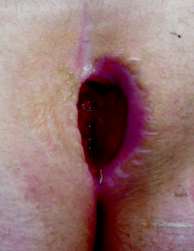
Fig. 52.6
An unhealed midline wound after wide excision
Treatment of Failures and Recurrences
NonSurgical Treatment
Some recurrences are fairly trivial and cause little upset to the patient. Occasional dressings and antibiotics may be preferable to more surgery. Shaving may keep some of the symptoms at bay. This option should be considered if the alternative is to offer a more extensive procedure with the risk of subsequent failure.
Simple Laying Open
Minimal disease with adequate marsupialization will solve a number of minor failures. This is performed frequently by many surgeons and probably goes unreported. It may be conducted in the out-patient clinic.
Wide Excision With or Without Primary Closure
Wide excision should not be performed because many failures occur after this technique and the unhealed midline wound is a debilitating consequence. To repeat the operation with a risk of a larger unhealed wound is not acceptable today when off-midline closure clearly has been shown to be superior [4 5]. The risk of failure will be similar to the risk after primary surgery.
Off-Midline Skin Closure
Karydakis’ operation may be performed for recurrent disease or failures. The success of this operation is similar to when it is carried out for primary disease and is a good option. It remains, however, a fairly extensive procedure and has been superseded by modifications. Bascom’s cleft closure or cleft lift achieves a similar outcome. It is perfectly suited to the unhealed midline wound and results in rapid and successful healing. In a series of 150 patients [80] in whom Bascom’s cleft closure or lift procedures were performed in 63 % for recurrent disease or for the unhealed midline wound, there was a 96 % success rate for healing. No patients had major breakdown of their wound, and only 5 % recurred requiring further surgery.
More Complex Skin Flap Procedures
These may also be performed for failures and recurrent disease. They are more suited to this situation rather than for primary disease unless the latter is very extensive. They are more appropriately used after failure of a Karydakis or cleft lift procedure.
Conclusion
In patients with minimal symptoms from pilonidal disease and those having drainage of a single acute abscess, there may not need to be any intervention and cases can be managed expectantly. Nonsurgical treatments of pilonidal disease such as phenol injections and simple shaving with careful natal cleft hygiene may be of value, but their long-term results are not known. Complications of treatment are most likely to be avoided, although recurrence rates may be high.
Simple disease requires simple surgery. Pit picking using Bascom’s technique has good results, and coring out the pits with a trephine may also be performed and repeated. More complex or recurrent disease with unhealed midline wounds can be successfully treated using modified Karydakis procedures such as the Bascom cleft lift operation, which avoids wide excision of the abscess, flattens the cleft, and is increasingly performed nowadays as a day case procedure under local anaesthesia, with good long-term outcomes and excellent cosmetic results. Z and V-Y flap procedures as well as the Limberg flap after wide excision of the abscess have been successful in this situation but they require general anesthesia, are more difficult to conduct as outpatient procedures, and failures may be hard to correct. Their cosmetic outcome may also be unsatisfactory. These are best reserved for situations where the cleft lift has failed and perhaps in extensive recurrent bilateral disease.
The success of nonexcisional surgery and the theory that midline skin pits are the cause of pilonidal disease means that there is now no rational basis or need for excision of the abscess in pilonidal disease. However, the major benefit of these newer simpler procedures is that they do not cause the occasional debilitating complications of pilonidal surgery by avoiding long sutured or unsutured midline wounds deep in the natal cleft. These days, many of the complications of treatment of pilonidal sinus disease can be prevented. Patients should be informed about these problems and alternative treatment regimes should be discussed. Surgeons who do not have a particular interest in the condition should avoid treating these patients because the consequences of failure can be quite devastating. It is helpful and gratifying to know that, should they occur, most failures and complications can be treated with good outcomes. There is no place, therefore, for allowing patients with poor outcomes to continue with unhealed wounds and infections indefinitely.
References
3.
4.
5.
6.
No Authors Listed. Pilonidal sinus: is surgery alone enough? Colorectal Dis. 2003; 5:205.
8.
9.
11.


