Reversible
Irreversible
Technical problems
Vascular disease
Microsurgical techniques
Atherosclerosis
Reduced blood flow
Radiation injury
Postoperative management/anticoagulation
Malformations
Patient positioning
Hypercoagulability
Delay in diagnosis
Systemic disease
Delay in salvage
39.3.1 Reversible Causes Due to Outer Circumstances
39.3.1.1 Microsurgical Technique and Intraoperative Flap Handling
The initial thought after failure of a free flap procedure will generally lead a self-critical microsurgeon to investigate whether technical reasons are directly responsible for the flap loss [8]. Indeed, many flap losses are attributable to inadequate surgical technique and planning [9, 10]. It is also well known that around 90 % of anastomotic thromboses occur in the venous part and that therefore the venous part has to be considered as the most frequent cause of anastomotic failure [11]. Therefore, immediate revision is mandatory when there is any suspicion of thrombosis postoperatively [12, 13]. Using coupler systems for venous anastomosis is believed to lower the complication rate on the venous side [14].
Risk of vessel injury and increased endothelial lesion followed by excessive platelet adhesion can be minimized by meticulous technique [15].
This includes avoiding to grasp the intima unduly with sharp instruments, avoiding tension at the anastomosis site, or undue clamping of vessels resulting in inner intimal lesions, not directly visible from the outside. It is advisable that if in doubt, vascular interpositional grafts should be used. Size match of flap vessel and recipient vessel should be ensured to avoid blood flow disturbances. At flap inset, tension on and kinking of the pedicle should be avoided [16]. In our own clinical practice, we found it helpful to protect the microanastomosis from kinking and from direct compression from the outside (such as by hematoma or seroma formation) by covering the anastomotic site with fibrin sealant [2, 17].
39.3.1.2 Postoperative Management
Careful positioning of the patient to prevent compression of the pedicle or the flap itself is crucial for successful outcome of free flap transfers, e.g., in the lower extremity or in the perineal or sacral region.
If in free muscle flaps skin pedicles are used, one has to make sure that the monitor island is sufficiently perfused and reflects the perfusion status of the muscles itself to rule out a false decision making [18].
The use of systemic anticoagulation varies widely, but routine use is not uniformly agreed upon by all microsurgeons [19, 20]. Dextran, aspirin, heparin, and low-molecular-weight heparins are the most commonly used pharmacologic agents. Particular indications such as bypass free flaps have been the subject of a more aggressive anticoagulative regimen. Given that evidence-based recommendations are not available thus far, the German-Speaking Society for Microsurgery has recently published its attempts to formulate potential guidelines for anticoagulation regimen after microsurgical free flap transfer [21].
When a second microvascular free flap is going to be performed to achieve a reconstructive goal following flap failure, systemic anticoagulation is often applied if risk factors persist as unfavorable conditions that are not correctable such as radiation tissue injury or radionecrosis [22]. This holds also true for complex microsurgical procedures including arterial bypasses or arteriovenous loops between flap and recipient site. In such interdisciplinary cases in our institution, we apply oral medication for systemic anticoagulation for half a year.
39.3.1.3 Patient-Inherent Irreversible Causes
These issues need to be considered in particular when evaluating the reconstructive options after free flap loss since they cannot be altered and may pose a limiting factor to secondary free flap reconstruction that has not been considered thoroughly enough at initial evaluation.
39.3.1.4 Vascular Disease
Atherosclerosis is one major threat to successful microsurgical free flap reconstruction. In particular, recipient vessels in the head and neck are and in the lower extremity may often be of poor quality, increasing the risk of anastomosis-related complications, embolism or thrombosis formation. When there is peripheral vascular disease of the lower extremity, free flap surgery itself may threaten survival of an already hypoperfused limb, necessitating the use of combined approaches using a bypass or vessel loop to provide both sufficient recipient vessels and secure perfusion of the compromised lower extremity [23, 24].
A history of diabetes and radiation injury may also severely compromise the quality of flap or recipient site vessels [25], the latter being of particular importance and frequently encountered in autologous breast reconstruction and sarcoma patients. When free flaps are buried, PET scanning may be helpful to determine the necessary measures [26] when Doppler or duplex sonography is not sufficient.
39.3.1.5 Systemic Disease
It is not uncommon that a failed free flap worsens the patient’s general condition, giving the patient an even greater risk when secondary free flap reconstruction is considered [27].
Clotting disorders including factor V Leiden, protein C and S deficiencies, and other conditions are not common, but an important cause to consider and identify before further efforts are undertaken to start another reconstructive effort using microsurgery [28, 29].
Myocardial infarction, stroke, respiratory failure, and morbid obesity and malnutrition are other severe conditions possibly precluding microsurgical reconstruction.
In each single case, the reconstructive goals must be reevaluated and potential new factors included in the analysis of patients after free flap failure.
39.4 Reevaluation of Reconstructive Goals
After a thorough assessment of the clinical course has been carried out and the cause for failure of the free flap procedure has been identified, a careful reevaluation of the status quo and the reconstructive goals is warranted.
The following factors must now be considered before the next steps towards a secondary reconstruction are being made, in particular if a second attempt for free flap microsurgical reconstruction is considered: first, the possible limitation of donor sites; second, the new defect that may be potentially larger than the first defect; and third, previously available recipient vessels that may no longer be available. Taking all these factors together, a second free flap may be more technically challenging, increasing the risk for another free flap failure in the same patient. The need and urgence to reach a reconstructive goal by the use of a free flap may vary considerably, reaching from indications mainly for restoration of aesthetic appearance (breast reconstruction, reanimation of the paralyzed face), functional reconstruction (reanimation of the paralyzed face), and limb salvage (lower leg reconstruction) to the essential coverage of vital structures such as major blood vessels [24] (Fig. 39.1).
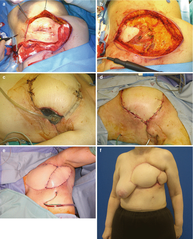
Fig. 39.1
(a) Palliative resection in fistulating inflammatory breast cancer after mastectomy and repeated irradiation (first cycle due to breast conserving therapy and second radiotherapy after mastectomy). (b) 43 cm × 27 cm large defect after palliative resection of fistulating inflammatory breast cancer. (c) After inset of extended DIEP flap to cover the large area, partial necrosis in zone IV occurred, necessitating debridement of the tip of the flap. We attempted wound conditioning with topical negative pressure therapy (TNP). (d) Secondary closure in the heavily irradiated field was attempted after wound conditioning with TNP. (e) Following wound breakdown after the secondary closure, a pedicled latissimus dorsi myocutaneous flap was necessary to achieve stable cover of the large wound area in the irradiated field. Note the fibrotic tissue changes and skin alteration below the latissimus dorsi flap in the upper abdominal wall soft tissue where the drain is placed through the skin. (f) Finally the wound was closed with conservative treatment of minor secondary healing in the lower wound margin between the latissimus dorsi and the DIEP flap within the irradiated area in this palliative situation rendering improved quality of life. The patient deceased 1 year later from pulmonary metastasis
39.4.1 Repeat Free Flap Procedure
Typical indications for where a repeat free flap procedure is recommended include the coverage of major vessels or vital structures, limb-threatening wounds in the extremity, and timely wound healing for potentially life-saving radiation. In such cases, it may be advisable to not risk another free flap but instead solve the problem with a pedicled flap to cover vital structures if possible [30–32].
Related posts:


Stay updated, free articles. Join our Telegram channel

Full access? Get Clinical Tree


