Management of Nerve Entrapment Syndromes

CHAPTER 21 Management of Nerve Entrapment Syndromes


NEURECTOMY AND NERVE BURIAL


I must state at the outset that I have not obtained good results over the decades with nerve surgery, regardless of the type and extent. Revision nerve surgery is worse, with unpredictable results, especially in patients with chronic pain syndromes, who are very difficult to treat. Moreover, I have seen numerous patients actually harmed by repeat nerve surgery performed with the best expectations. Accordingly, the need for caution in recommending nerve surgery for management of foot and ankle pain cannot be overemphasized.


An isolated neuroma of the sural nerve may be treated surgically in the appropriate patient. By this I mean a patient who does not have chronic pain and who has localized discomfort only, from pressure over the traumatized nerve. In general, the outcome with neurectomy and nerve burial seems to be better in patients in whom the nerve was scarred or cut as a result of previous surgery than in patients who present with isolated nerve pain after an injury. In the latter group of patients, the affected foot frequently is sensitive to pressure, light touch, compression, and nerve irritation. Many of these patients with chronic pain syndromes initially are treated with topical anesthetic medications and oral neuroleptic agents, and surgery often is undertaken as a last resort. In such cases, inadequacy of pharmacologic management or of the attempted surgical correction may be the beginning of a chronic pain syndrome, with never-ending problems. I therefore rarely perform these procedures unless absolutely necessary.


Neurectomy and nerve burial may be performed in conjunction with additional procedures for correction of midfoot or hindfoot deformity, the most common of which is a subtalar arthrodesis after a calcaneus fracture, when a sural neurectomy is performed. The incision varies according to the presence of previous incisions, the location of the neuroma, and the type of additional operation performed. From a vertical incision made posterior to the peroneal tendon sheath, the nerve is identified and inspected. The nerve must be traced from proximal to distal—that is, from the healthy to the abnormal portion of the nerve. Identifying a discrete neuroma is difficult because it usually is encased in scar. The neuroma frequently is a consequence of previous surgery and is not mobile, particularly if it is located under the earlier incision. The sural nerve should be traced more distally until either the scar or a definite neuroma is identified. The nerve is dissected further distally, and if no further continuity with the main body of the nerve is observed, it is transected, including the neuroma.


The nerve is now clamped and the tip of the nerve is cauterized either using the electrocautery or with phenol. More proximally, the nerve is passed with a clamp and a 4-0 suture deep to the peroneal tendons or musculature, depending on the level in the leg. Simple resection of the neuroma is never sufficient. Burial in a muscle may work, but the recurrence rate seems to be high, and burying the nerve in bone is preferable. The key is to obtain sufficient length on the exposed nerve so that with movement of the leg and contracture of the peroneal muscles, there is no tension on the nerve.


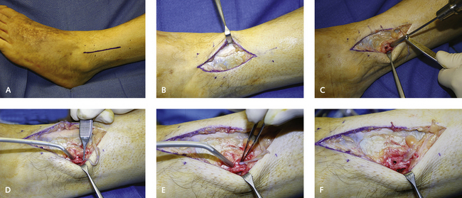
Burying the nerve in bone is difficult, because the nerve does not easily stay in position. Two small 2.5-mm unicortical drill holes are made through the fibula perpendicular to each other and about 1 cm apart. The nerve is positioned at one end of the bone tunnel, and the suction is applied to the other drill hole to draw the nerve deep into the hole in the fibula. This drill hole technique seems to be the most effective means of burying the nerve inside the fibula. Once the nerve has been passed into the fibular hole, the epineurium can be sutured onto the periosteum. The ankle should be taken through full dorsiflexion and plantar flexion to ensure that there is no traction on the nerve and that the nerve is freely mobile in the posterior aspect of the limb and encased in the fibula itself (Figure 21-1).


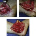
Occasionally, after certain types of injuries, including crush injuries, a neurectomy may not be necessary, and a nerve release after removal of the ligament or tendon may be sufficient (Figures 21-2 to 21-5

Only gold members can continue reading. Log In or Register to continue

Stay updated, free articles. Join our Telegram channel

Mar 6, 2016 | Posted by in Reconstructive surgery | Comments Off on Management of Nerve Entrapment Syndromes

Full access? Get Clinical Tree

Get Clinical Tree app for offline access