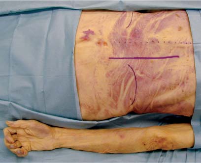
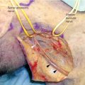
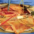
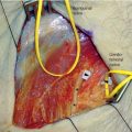
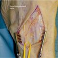
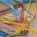
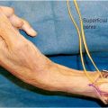
8 The lumbar plexus entails the ventral rami of the first three lumbar nerves, a contribution from the subcostal nerve, and the major portion of the fourth lumbar nerve. The plexus lies embedded within the mass of the psoas muscle and anterior to the lumbar vertebral spinous processes. The first lumbar spinal nerve receives a fascicle from the subcostal nerve and divides into upper and lower branches. The upper branch then splits into the iliohypogastric and ilioinguinal nerves, whereas the lower branch joins a fascicle from the second lumbar spinal nerve to become the genitofemoral nerve. The subcostal nerve, iliohypogastric nerve, ilioinguinal nerve, and genitofemoral nerve travel within the posterior wall of the abdomen and pelvis in a course parallel to one another. The iliohypogastric nerve gives off a lateral cutaneous branch to supply the skin on the anterolateral aspect of the buttock. It then courses superior to the inguinal canal and finally ends as the anterior cutaneous branch to the skin above the pubis. The ilioinguinal nerve pierces the internal oblique muscle above the anterior part of the iliac crest and then runs above and parallel to the inguinal ligament to traverse the inguinal canal. The nerve emerges through the external ring to eventually supply the skin over the root of the penis, the adjoining part of the femoral triangle, and the upper part of the scrotum (mons pubis and adjacent part of the labium majoris in the female). The genitofemoral nerve penetrates the psoas major muscle and divides into genital and femoral branches. The genital branch passes through the inguinal canal and supplies the cremaster muscle and the skin of the scrotum in the male and the round ligament in the female. The femoral branch supplies sensation to the skin of the upper part of the femoral triangle. The larger portion of the second lumbar spinal nerve, the entire third lumbar spinal nerve, and a portion from the fourth lumbar spinal nerve split into ventral (anterior) and dorsal (posterior) divisions. The ventral divisions unite to form the obturator nerve, and the dorsal divisions unite to form the femoral nerve (see Chapter 9). The obturator nerve has contributions from lumbar spinal nerves 2, 3, and 4. The contribution from the second lumbar spinal nerve is the smallest and it is frequently absent. The obturator supplies the obturator externus and the adductor muscles of the thigh, gives branches to the hip and knee joints, and contributes to the cutaneous innervation to the medial portion of the thigh. Once constituted from the lumbar spinal roots, the nerve descends through the psoas muscle parallel to the lumbosacral trunk. It emerges from the medial border of the psoas muscle approximately at the level of the pelvic brim. It travels medial to the sacroiliac joint downward over the sacral ala between the psoas muscle and the vertebral column. It moves into the lesser pelvis lying lateral to the ureter and internal iliac vessels. Joined by the obturator artery and vein, it then bends anteroinferiorly, lying on the obturator internus muscle to reach the obturator groove at the upper part of the obturator foramen. The nerve then passes through this groove to enter the thigh and there it divides into anterior and posterior branches. The two branches are separated first by the obturator externus muscle and then more inferiorly by the adductor brevis muscle. The anterior branch, as its name implies, runs anterior to the obturator externus and adductor brevis. It sends muscular branches to the adductor longus, adductor gracilis, and adductor brevis muscles. The anterior branch terminates as small cutaneous, vascular, and communicating branches. The cutaneous branch partially supplies innervation to the skin and fascia of the distal two thirds of the medial thigh. The posterior branch supplies the obturator externus muscle, the adductor magnus, and, inconstantly, the adductor brevis. It too ends as small vascular and joint capsule contributions. The lateral femoral cutaneous nerve is formed by contributions from the posterior divisions of the second and third lumbar spinal nerves (see Chapter 10). To expose the nerve roots that form the lumbar plexus and ~4 to 6 cm lateral to the neuroforamina a lateral extracavitary approach as seen in spine surgery may be utilized.1–3 This approach can be used in all regions of the lumbar and thoracic spine, but in the case of the lumbar plexus no higher than T12 is required. Exposure of the lower lumbar plexus requires resection of the dorsal ilium, and therefore a pelvic brim approach may be more useful if that part of the plexus is the focus (see later). For this exposure a three-quarter prone position is preferred for visualization as well as lessened blood loss secondary to reduced abdominal compression. A bolster is placed under the ipsilateral thorax and pelvis. The surgeon works from the ipsilateral side. A paramedian incision is laid out ~4 cm off the midline. It should extend from 3 to 4 cm below the level of the posterior iliac crest and proximally as required (Fig. 8-1). The skin and subcutaneous tissue are divided after being infiltrated with 1% lidocaine with epinephrine in a 1:100,000 concentration. Once the skin and subcutaneous tissue are incised the lumbodorsal fascia are visible (Fig. 8-2). This fascia should be incised to reveal the erector spinae muscle (medially) and the quadratus lumborum muscle (laterally). Probing along the muscle edge reveals a plane between the two muscles that can then be followed down to the transverse processes (Figs. 8-3 and 8-4). Once at the transverse processes, which will be palpable before they are visible, a subperiosteal dissection hugging the undersurface of the process takes the surgeon safely to the vertebral body and neural foramen, if necessary. The top of the processes may also be cleared of overlying soft tissue (Fig. 8-5). At this point the psoas muscle should be visible beneath the transverse processes. The processes may be carefully removed with a rongeur for enhanced visualization (Fig. 8-6). With gentle dissection the elements of the plexus, which are heading in an oblique direction laterally and inferiorly from the midline, become visible (Fig. 8-7).
LUMBAR PLEXUS
ANATOMY
POSITIONING AND SURGICAL EXPOSURE
Lateral Extracavitary Approach, for Posterior Proximal Exposure
Lumbar Plexus
Only gold members can continue reading. Log In or Register to continue

Full access? Get Clinical Tree


