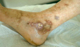
Fig. 26.1
Livedo

Fig. 26.2
Ulceration and livedoid aspect

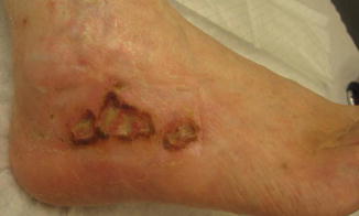
Fig. 26.3
Chronic ulcerations

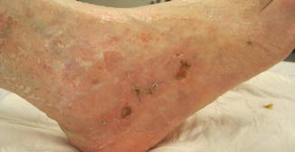
Fig. 26.4
Healing with white atrophic surrounding plaques and telangiectases
References
1.
Khenifer S, Thomas L, Balme B, Delle S. Livedoid vasculopathy: thrombotic or inflammatory disease ? Clin Exp Dermatol. 2009;35:693–8.CrossRef
2.
Hairston B, Davis MD, Pittelkow MR, Ahmed I. Livedoid vasculopathy: further evidence for procoagulant pathogenesis. Arch Dermatol. 2006;142(11):1413–8.CrossRefPubMed
Stay updated, free articles. Join our Telegram channel

Full access? Get Clinical Tree


